Chapter: Medical Physiology: Somatic Sensations: I. General Organization, the Tactile and Position Senses
Somatosensory Cortex
Somatosensory Cortex
Before discussing the role of the cerebral cortex in somatic sensation, we need to give an orientation to the various areas of the cortex. Figure 47–5 is a map of the human cerebral cortex, showing that it is divided into about 50 distinct areas called Brodmann’s areasbased on histological structural differences. This map is important because virtually all neurophysiologists and neurologists use it to refer by number to many of the different functional areas of the human cortex.
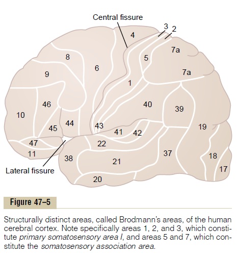
Note in the figure the large central fissure (also called central sulcus) that extends horizontally across the brain. In general, sensory signals from all modali-ties of sensation terminate in the cerebral cortex immediately posterior to the central fissure. And, generally, the anterior half of the parietal lobe is concerned almost entirely with reception and inter-pretation of somatosensory signals. But the posterior half of the parietal lobe provides still higher levels of interpretation.
Visual signals terminate in the occipital lobe, and auditory signals in the temporal lobe.
Conversely, that portion of the cerebral cortex ante-rior to the central fissure and constituting the poste-rior half of the frontal lobe is called the motor cortex and is devoted almost entirely to control of muscle contractions and body movements. A major share of this motor control is in response to somatosensory signals received from the sensory portions of the cortex, which keep the motor cortex informed at each instant about the positions and motions of the differ-ent body parts.
Somatosensory Areas I and II. Figure 47–6 shows two sep-arate sensory areas in the anterior parietal lobe called somatosensory area Iand somatosensory area II. Thereason for this division into two areas is that a distinct and separate spatial orientation of the different parts of the body is found in each of these two areas. However, somatosensory area I is so much more extensive and so much more important than somatosensory area II that in popular usage, the term “somatosensory cortex” almost always means area I.
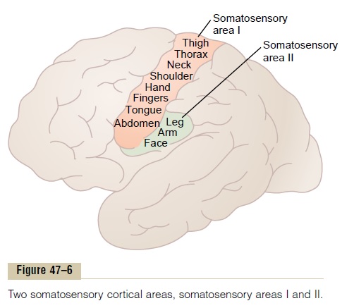
Somatosensory area I has a high degree of localiza-tion of the different parts of the body, as shown by the names of virtually all parts of the body in Figure 47–6. By contrast, localization is poor in somatosensory area II, although roughly, the face is represented anteriorly, the arms centrally, and the legs posteriorly.
Little is known about the function of somatosensory area II. It is known that signals enter this area from the brain stem, transmitted upward from both sides of the body. In addition, many signals come secondarily from somatosensory area I as well as from other sensory areas of the brain, even from the visual and auditory areas. Projections from somatosensory area I are required for function of somatosensory area II. However, removal of parts of somatosensory area II has no apparent effect on the response of neurons in somatosensory area I. Thus, much of what we know about somatic sensation appears to be explained by the functions of somatosensory area I.
Spatial Orientation of Signals from Different Parts of the Body in Somatosensory Area I. Somatosensory area I liesimmediately behind the central fissure, located in the postcentral gyrus of the human cerebral cortex (in Brodmann’s areas 3, 1, and 2).
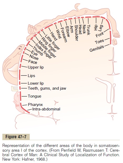
Figure 47–7 shows a cross section through the brain at the level of the postcentral gyrus, demonstrating representations of the different parts of the body in separate regions of somatosensory area I. Note, however, that each lateral side of the cortex receives sensory information almost exclusively from the oppo-site side of the body.
Some areas of the body are represented by large areas in the somatic cortex—the lips the greatest of all, followed by the face and thumb—whereas the trunk and lower part of the body are represented by rela-tively small areas. The sizes of these areas are directly proportional to the number of specialized sensory receptors in each respective peripheral area of the body. For instance, a great number of specialized nerve endings are found in the lips and thumb, whereas only a few are present in the skin of the body trunk.
Note also that the head is represented in the most lateral portion of somatosensory area I, and the lower part of the body is represented medially.
Layers of the Somatosensory Cortex and Their Function
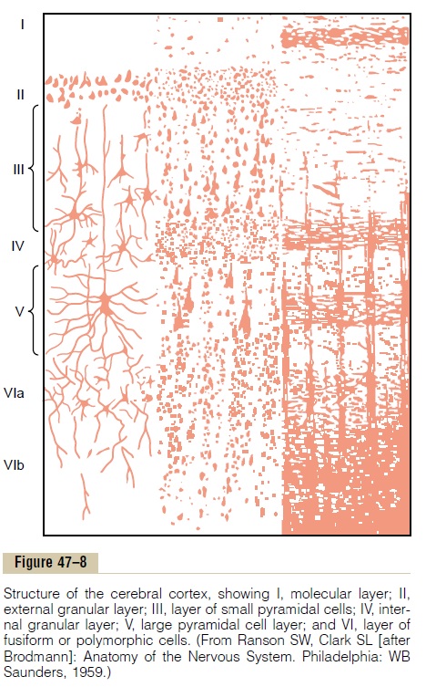
The cerebral cortex contains six layers of neurons, beginning with layer I next to the brain surface and extending progressively deeper to layer VI, shown in Figure 47–8. As would be expected, the neurons in each layer perform functions different from those in other layers. Some of these functions are:
1.The incoming sensory signal excites neuronal layer IV first; then the signal spreads toward the surface of the cortex and also toward deeper layers.
2.Layers I and II receive diffuse, nonspecific input signals from lower brain centers that facilitate specific regions of the cortex;. This input mainly controls the overall level of excitability of the respective regions stimulated.
3.The neurons in layers II and III send axons to related portions of the cerebral cortex on the opposite side of the brain through thecorpuscallosum.
4.The neurons in layers V and VI send axons to the deeper parts of the nervous system. Those in layer V are generally larger and project to more distant areas, such as to the basal ganglia, brain stem, and spinal cord where they control signal transmission. From layer VI, especially large numbers of axons extend to the thalamus, providing signals from the cerebral cortex that interact with and help to control the excitatory levels of incoming sensory signals entering the thalamus.
The Sensory Cortex Is Organized in Vertical Columns of Neurons; Each Column Detects a Different Sensory Spot on the Body with a Specific Sensory Modality
Functionally, the neurons of the somatosensory cortex are arranged in vertical columns extending all the way through the six layers of the cortex, each column having a diameter of 0.3 to 0.5 millimeter and con-taining perhaps 10,000 neuronal cell bodies. Each of these columns serves a single specific sensory modal-ity, some columns responding to stretch receptors around joints, some to stimulation of tactile hairs, others to discrete localized pressure points on the skin, and so forth. At layer IV, where the input sensory signals first enter the cortex, the columns of neurons function almost entirely separately from one another. At other levels of the columns, interactions occur that initiate analysis of the meanings of the sensory signals.
In the most anterior 5 to 10 millimeters of the post-central gyrus, located deep in the central fissure in Brodmann’s area 3a, an especially large share of the vertical columns respond to muscle, tendon, and joint stretch receptors. Many of the signals from these sensory columns then spread anteriorly, directly to the motor cortex located immediately forward of the central fissure. These signals play a major role in controlling the effluent motor signals that activate sequences of muscle contraction.
As one moves posteriorly in somatosensory area I, more and more of the vertical columns respond to slowly adapting cutaneous receptors, and then still farther posteriorly, greater numbers of the columns are sensitive to deep pressure.
In the most posterior portion of somatosensory area I, about 6 per cent of the vertical columns respond only when a stimulus moves across the skin in a particular direction. Thus, this is a still higher order of interpre-tation of sensory signals; the process becomes even more complex as the signals spread farther backward from somatosensory area I into the parietal cortex, an area called thesomatosensory association area, as we discuss subsequently.
Functions of Somatosensory Area I
Widespread bilateral excision of somatosensory area I causes loss of the following types of sensory judgment:
1.The person is unable to localize discretely the different sensations in the different parts of the body. However, he or she can localize these sensations crudely, such as to a particular hand, to a major level of the body trunk, or to one of the legs. Thus, it is clear that the brain stem, thalamus, or parts of the cerebral cortex not normally considered to be concerned with somatic sensations can perform some degree of localization.
2.The person is unable to judge critical degrees of pressure against the body.
3.The person is unable to judge the weights of objects.
4.The person is unable to judge shapes or forms of objects. This is called astereognosis.
5.The person is unable to judge texture of materials because this type of judgment depends on highly critical sensations caused by movement of the fingers over the surface to be judged.
Note that in the list nothing has been said about loss of pain and temperature sense. In specific absence of only somatosensory area I, appreciation of these sensory modalities is still preserved both in quality and intensity. But the sensations are poorly localized, indi-cating that pain and temperature localization depend greatly on the topographical map of the body in somatosensory area I to localize the source.
Related Topics