Chapter: The Massage Connection ANATOMY AND PHYSIOLOGY : Muscular System
Sliding Filament Mechanism
SLIDING FILAMENT MECHANISM
The sliding filament mechanism explains the process of muscle contraction at the molecular level. This process is initiated by impulses from the nerve that innervates the muscle fiber.
Muscle-Nerve Communication
Skeletal muscle only contracts when stimulated by the communicating nerve. Each muscle fiber is in contact with a nerve ending. The cell body of the nerve fiber (a single neuron) is located in the spinal cord, brainstem, or brain, according to where the skeletal muscle is located and to where it originated in the embryonic stage. The axons of these neurons extend from the cell bodies to individual muscles.
For example, when we say that the ulna nerve sup-plies the muscles of the thumb, we are indicating the bundles of axons of motor neurons that go together as the ulna nerve before they split to supply the indi-vidual muscle fibers of muscles that move the thumb.
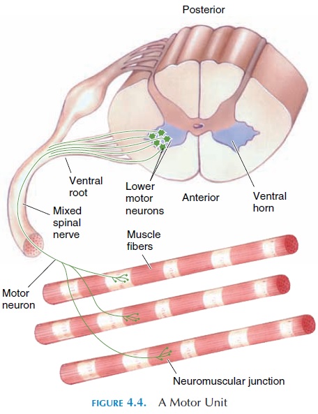
The axons branch when they reach the muscles they supply, and each axon communicates with one or more muscle fibers. Thus, if a neuron is stimu-lated, all of the muscle fibers it communicates with will contract. The axon, its branches, and all the mus-cle fibers it supplies are known as a motor unit (see Figure 4.4). A motor neuron innervates an average of 150 muscle fibers. However, in muscles that require precise control, a neuron may innervate only two or three fibers.
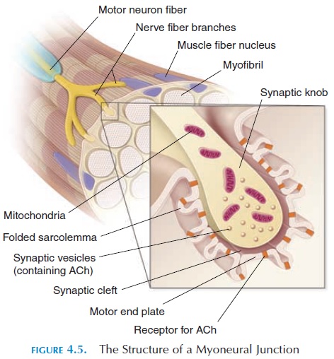
At the point where they come in close contact with the muscle fiber, each nerve ending is modified. The region where the nerve and the muscle communicates is themyoneural junction or neuromuscular junc-tion (see Figure 4.5). The nerve ending expands hereto form a synaptic knob. The cytoplasm of the nerve ending has vesicles containing molecules of acetyl-choline (ACh). A small gap—synaptic cleft—existsbetween the synaptic knob and the sarcolemma of the muscle fiber. The portion of the sarcolemma directly under the synaptic knob is the motor endplate. The sarcolemma underlying the synaptic knob has proteins (receptors) on its surface that have an affinity for ACh.
The receptors are actually ion channels that are regulated by ACh. The connective tissue matrix in the synaptic cleft has acetylcholinesterase enzymes that can destroy ACh.
Nerve Impulse and Activityin the Myoneural Junction
When a specific muscle is moved, nerve impulses or action potentials pass down the nerve axon until the myo-neural junction is reached. This triggers opening of calcium channels in the nerve axon, with resultant movement of calcium into the axon. The calcium movement triggers vesicles containing ACh to fuse with the nerve cell membrane and release ACh into the synaptic cleft. ACh attaches to the ACh receptors on the motor endplate, resulting in opening of the ion channels in the sarcolemma. The changes produced by ACh only last for a short time because the acetyl-cholinesterase located in the synaptic cleft begins to break down ACh. The sarcolemmal properties reach that of the resting stage when all ACh is destroyed.
Excitation-Contraction Coupling
When ACh binds to the receptor, the change that oc-curs is the opening of sodium channels on the sar-colemma. This results in sodium (which is of a higher concentration outside the cell than inside) rushing into the sarcoplasm of the muscle fiber. At rest, the in-side of the muscle fiber is electrically negative com-pared to the outside. When positively charged sodium enters the cell, the inside becomes positive. This change in potential triggers a series of reactions in-side the muscle fiber at the molecular level that pro-duces muscle contraction (see Figure 4.6). The link between the potential change in the sarcolemma and the contraction of the muscle is known as excitation-contraction coupling.
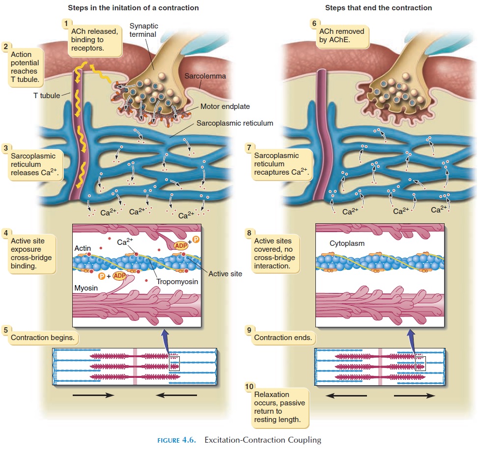
The potential change at the sarcolemma continues down into the T tubules, directly into the muscle fiber where it triggers the sarcoplasmic reticulum to re-lease calcium into the sarcoplasm. The calcium binds to the calcium site on the troponin (the protein on the actin). This binding causes the troponin to shift the tropomyosin, exposing the active site for myosin lo-cated on actin. When exposed by the movement of tropomyosin, the myosin heads attach to the active site.
The myosin head moves toward the M line in a hingelike action, deriving energy from breaking down ATP (adenosine triphosphate).
ATP → ADP (adenosine diphosphate) phosphate
ADP and phosphate, the breakdown products, move away and another ATP binds to the myosin head to provide energy. The attachment of the next ATP to the myosin head causes the myosin to detach from the actin site, move back into its original posi
Thus, the actin is moved closer to the M line, with the myosin attaching and detaching from the active sites on subsequent actin molecules. This process contin- ues for as long as calcium ions are present in the sar-coplasm and ATP is available for energy. Thus, the actin filament slides between the myosin filaments, shortening the muscle fiber.
Relaxation of Muscle Fiber
Soon after the impulse arrives, the sarcoplasmic reticulum that released its calcium content into the sarcoplasm starts pumping the calcium back, using ATP as energy. If no other impulse arrives, the cal-cium continues to be pumped back from the sar-coplasm until the resting levels of calcium are reached. When the calcium level drops in the sar- coplasm, the troponin loses calcium from its binding site and the tropomyosin returns to its original posi-tion, blocking the active sites in the actin and ending the actin-myosin interaction. This results in relax-ation of the muscle.
As long as action potential/impulses arrive at the myoneural junction, ACh continues to be released. As mentioned, ACh is broken down by acetylcholines-terase. If impulses stop, no additional ACh is released, and the remaining ACh in the synaptic cleft is removed by acetylcholinesterase. The sodium channels that were opened by ACh binding to receptors close, and the po-tential inside the sarcoplasm is brought back to its rest-ing state. (The details of how the potential is brought back are not elaborated here).
Time Lapse Between NerveImpulse and Contraction
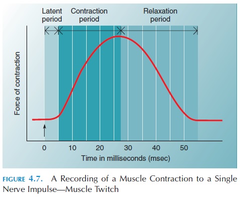
The latent period is the short duration of time that elapses when a muscle responds to a single impulse before the muscle begins to shorten. This includes the time taken for the impulse to travel down the nerve, release ACh, and all the reactions that take place within the sarcoplasm before sliding of the fila-ments occurs. In addition, it includes the time for the tendon and other connective tissue (e.g., perimysium, epimysium) to be stretched before the force can be transmitted to the bone. Because the muscle is at-tached to the bone via the connective tissue tendon, the tendon (with its elastic fibers) must be stretched for the force produced by the muscle to reach the bone. This is similar to lifting a ball tied to an elastic band. Before the ball can be lifted off the ground, the elastic band must be tautly stretched. In the muscle, the first few impulses produce enough muscle con-traction to stretch the tendon. If impulses continue to come down the nerve, the tension is transmitted to the bone more effectively.
The contraction period is the duration of muscle contraction in response to a nerve impulse, and the relaxation period is the duration taken by the fiberto relax after a contraction (see Figure 4.7). The recording of the response of the muscle to a single nerve impulse is known as a muscle twitch. For a short time after the first impulse arrives, the muscle is unable to respond to a second stimuli. This period is known as the refractory period. The refractory pe-riod in skeletal muscle is short, about 5 milliseconds (ms). As a result of the short refractory period, an-other impulse arriving just after 5 ms can produce a muscle response. Note that this impulse would arrive during the contraction period of the first muscle twitch. Therefore, it is possible for the response to the second impulse to fuse with that of the first to produce a sustained contraction.
Fortunately, the refractory period in cardiac mus-cle is long, about 300 ms. This prevents sustained contractions of the heart—a situation that would stop circulation of blood.
Related Topics