Chapter: Human Neuroanatomy(Fundamental and Clinical): Internal Structure of the Spinal Cord
Myotatic or Stretch Reflexes
Myotatic or Stretch Reflexes
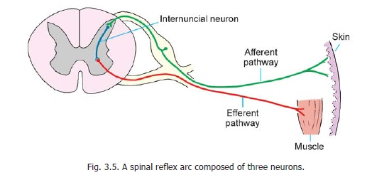
Sudden stretching of a muscle (by tapping its tendon) produces reflex contraction of the muscle. The pathway for this reflex involves two neurons only. Stretching of the muscle stimulates proprioceptive nerve endings located in muscle spindles and other receptors. These impulses are carried to the spinal cord by neurons that synapse with motor neurons in the ventral grey columns (Fig. 3.5). Fibres arising from these motor neurons reach the muscle and produce contraction. Stretch reflexes are abolished if any part of the pathway for it (i.e., the reflex arc) is interrupted. Under certain conditions these reflexes may be exaggerated. From a clinical point of view it is important to know the level of the spinal cord at which each reflex is mediated. Some of the important stretch reflexes are described below. The spinal segments concerned are given in brackets.
1. The knee jerk or patellar tendon reflex consists of extension of the leg by contraction of the quadriceps when the ligamentum patellae is tapped (L2, L3, L4).
2. The ankle jerk or Achilles tendon reflex consists of plantar flexion of the foot on tapping the tendo calcaneus (L5, S1, S2).
3. The biceps tendon reflex consists of flexion of the forearm on tapping the biceps tendon (C5, C6).
4. The triceps tendon reflex consists of extension of the forearm on tapping the triceps tendon (C6, C7, C8).
5. The supinator jerk (or radial periosteal reflex) consists of flexion of the forearm when the distal end of the radius is tapped (C7, C8). Note that the muscle responsible for this reflex is the brachioradialis, not the supinator. It is called the supinator jerk because the brachioradialis was at one time called the supinator longus. This is a periosteal reflex, not a tendon reflex. According to some authorities the spinal segments responsible for the reflex are C5, 6,7.
6. The wrist tendon reflex consists of flexion of the fingers on percussion on wrist tendons (C8, T1).
7. The jaw or masseter reflex is a myotatic reflex mediated through the trigeminal nerve (and not through the spinal cord). To elicit this reflex the patient is asked to open the mouth slightly. The examiner places his index finger over the middle of the patient’s chin and taps it. This results in bilateral contraction of the masseter and temporalis muscles. Both afferent and efferent components of the reflex arc pass through the mandibular division of the trigeminal nerve, the nuclei concerned being located in the pons.
Related Topics