Chapter: Medical Physiology: Cortical and Brain Stem Control of Motor Function
Motor Cortex and Corticospinal Tract
MOTOR CORTEX AND CORTICOSPINAL TRACT
Figure 55–1 shows the functional areas of the cerebral cortex. Anterior to the central cortical sulcus, occupying approximately the posterior one third of the frontal lobes, is the motor cortex. Posterior to the central sulcus is the somatosensory cortex, whichfeeds the motor cortex many of the signals that initiate motor activities.
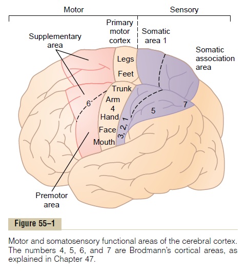
The motor cortex itself is divided into three subareas, each of which has its own topographical representation of muscle groups and specific motor func-tions: (1) theprimary motor cortex, (2) the premotor area, and (3) the supple-mentary motor area.
Primary Motor Cortex
The primary motor cortex, shown in Figure 55–1, lies in the first convolution of the frontal lobes anterior to the central sulcus. It begins laterally in the sylvian fissure, spreads superiorly to the uppermost portion of the brain, and then dips deep into the longitudinal fissure. (This area is the same as area 4 in Brodmann’s classification of the brain cortical areas, shown in Figure 47–5.)
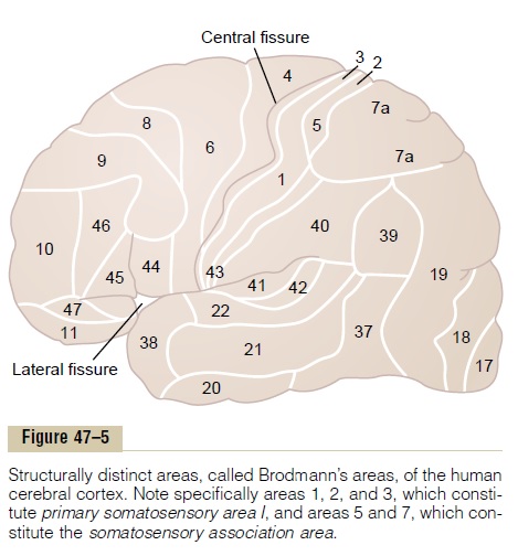
Figure 55–1 lists the approximate topographical representations of the dif-ferent muscle areas of the body in the primary motor cortex, beginning with the face and mouth region near the sylvian fissure; the arm and hand area, in the midportion of the primary motor cortex; the trunk, near the apex of the brain; and the leg and foot areas, in the part of the primary motor cortex that dips into the longitudinal fissure. This topographical organization is demonstrated even more graphically in Figure 55–2, which shows the degrees of representation of the different muscle areas as mapped by Penfield and Rasmussen. This mapping was done by electrically stimulating the different areas of the motor cortex in human beings who were undergoing neurosurgical operations. Note that more than one half of the entire primary motor cortex is concerned with controlling the muscles of the hands and the muscles of speech. Point stimulation in these hand and speech motor areas on rare occasion causes contraction of a single
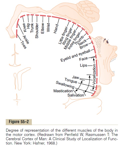
muscle; most often, stimulation contracts a group of muscles instead. To express this in another way, exci-tation of a single motor cortex neuron usually excites a specific movement rather than one specific muscle. To do this, it excites a “pattern” of separate muscles, each of which contributes its own direction and strength of muscle movement.
Premotor Area
The premotor area, also shown in Figure 55–1, lies 1 to 3 centimeters anterior to the primary motor cortex, extending inferiorly into the sylvian fissure and supe-riorly into the longitudinal fissure, where it abuts the supplementary motor area, which has functions similar to those of the premotor area. The topograph-ical organization of the premotor cortex is roughly the same as that of the primary motor cortex, with the mouth and face areas located most laterally; as one moves upward, the hand, arm, trunk, and leg areas are encountered.
Nerve signals generated in the premotor area cause much more complex “patterns” of movement than the discrete patterns generated in the primary motor cortex. For instance, the pattern may be to position the shoulders and arms so that the hands are properly oriented to perform specific tasks. To achieve these results, the most anterior part of the premotor area first develops a “motor image” of the total muscle movement that is to be performed. Then, in the poste-rior premotor cortex, this image excites each succes-sive pattern of muscle activity required to achieve the image.This posterior part of the premotor cortex sends its signals either directly to the primary motor cortex to excite specific muscles or, often, by way of the basal ganglia and thalamus back to the primary motor cortex. Thus, the premotor cortex, basal ganglia, thal-amus, and primary motor cortex constitute a complex overall system for the control of complex patterns of coordinated muscle activity.
Supplementary Motor Area
The supplementary motor area has yet another topo-graphical organization for the control of motor func-tion. It lies mainly in the longitudinal fissure but extends a few centimeters onto the superior frontal cortex. Contractions elicited by stimulating this area are often bilateral rather than unilateral. For instance, stimulation frequently leads to bilateral grasping movements of both hands simultaneously; these movements are perhaps rudiments of the hand func-tions required for climbing. In general, this area func-tions in concert with the premotor area to provide body-wide attitudinal movements, fixation movements of the different segments of the body, positional movements of the head and eyes, and so forth, as back-ground for the finer motor control of the arms and hands by the premotor area and primary motor cortex.
Related Topics