Chapter: Microbiology and Immunology: Morphology and Physiology of Bacteria
Light microscopy
◗ Light microscopy
Light microscopy, as the name suggests, uses natural or artificial transmitted light as the source of light. Resolving power of microscope is an important component of light microscopy. It is the ability of the lens system to distinguish two closely placed objects as distinct and separate entities. It is dependent on the wavelength of light used to illuminate the object and on the numerical aperture of the microscope. It is about half of the wavelength of light being used. For example, the smallest particle which can be resolved by yellow light with a wavelength of 0.4 mm is about 0.2 mm. Proper use of condenser that focuses light on the plane of the object facilitates optimization of the resolving power of the microscope. Resolving power of the microscope is enhanced further by adjusting the medium through which light passes between the object and objective lens. The use of immersion oil, whose refractive index is similar to that of the glass, improves the resolution of the microscope. The numerical aperture of the microscope is defined as the light gathering power of the microscope. Different types of light microscopy include (a) bright-field microscopy, (b) dark-ground microscopy, (c) phase-contrast microscopy, and (d) interference microscopy.
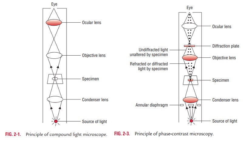
1. Bright-field microscopy: Bright-field microscopy (alwaysreferred to as ordinary light microscopy) is the most common form of light microscopy that uses a compound light micro-scope. A compound light microscope primarily consists of a compound lens system that contains a number of objective lenses, such as lenses of low power (310), high power (340), and oil immersion (3100). It also contains a fixed ocular (eye piece) lens, usually of 310 or 35. Final magnification of an object is the multiplication of lens power of the objective with that of the eye piece (Fig. 2-1). The bright-field microscopy has many uses.
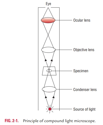
It may be used to examine either wet films or “hanging drop” for demonstration of the motility of flagellated bacteria (e.g., Escherichia coli, Pseudomonas aeruginosa, etc.) and protozoa (e.g., Trichomonasvaginalis, Giardia intestinalis, etc.). The wet prep-aration is also useful for demonstration of microorganisms in urine or feces, and also for detection of fungi in the skin.It is useful for demonstration of the structural details.
It is also useful for measuring approximate size of the bacte-ria, fungi, and protozoa in stained preparations.
2. Dark-ground microscopy: The dark-ground microscopymakes use of dark-ground microscope, a special type of compound light microscope. The dark-field condenser with a central circular stop, which illuminates the object with a cone of light, is the most essential part of the dark-ground microscope. This microscope uses reflected light instead of transmitted light used in the ordinary light microscope (Fig. 2-2). It prevents light from falling directly on the objective lens. Light rays falling on the object are reflected or scattered onto the objective lens with the result that the microorganisms
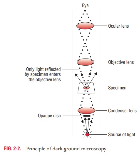
· It is useful for demonstration of very thin bacteria (such as, spirochetes) not visible under ordinary illumination, since the reflection of the light makes them appear larger. This is a frequently used method for rapid demonstration of Treponemapallidumin clinical specimens.
· It is also useful for demonstration of motility of flagellated bacteria and protozoa.
3. Phase-contrast microscopy: Phase-contrast microscopymakes use of a specific optical system that converts differences
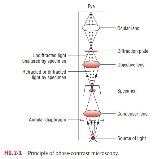
in phase in an organism into differences in intensity of light thereby producing light and dark contrast in the image (Fig. 2-3).
The optical system includes a special condenser and objec-tive lens which can be fitted to an ordinary light microscope to convert it into a phase-contrast microscope. The phase-con-trast microscopy has following uses:
o It is immensely useful for examination of living micro-organisms particularly protozoa (e.g., T. vaginalis, Entamoebahistolytica, etc.)
o It is useful for examining the internal structures of a living cell by improving the contrast and differentiating structures within the cell that differs in their thickness and refractive index.
4. Interference microscopy: This is another specialized application of light microscopy used for demonstrating cell organ-elles. It is also useful for quantitative measurement of the chemical constituents of the cells, such as proteins, lipids, and nucleic acids.
Related Topics