Chapter: Essentials of Anatomy and Physiology: Reproductive System
Formation of Gametes
FORMATION OF GAMETES
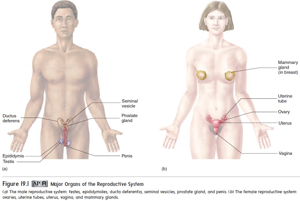
The testes in males and the ovaries in females (figure 19.1) pro-duce gametes (gam′ ētz), or sex cells. The formation of gametes in males and females occurs by a type of cell division called meiosis (mı̄-ō′ sis; a lessening) .
Meiosis occurs only in the testis and ovary. During meiosis, one cell undergoes two consecutive cell divisions to produce four daugh-ter cells, each having half as many chromosomes as the parent cell.
The two divisions of meiosis are called meiosis I and meiosis II. Like mitosis, each division of meiosis has prophase, metaphase, anaphase, and telophase. However, there are distinct differences between meiosis and mitosis.
Before meiosis begins, all the chromosomes are duplicated. At the beginning of meiosis, each of the 46 chromosomes consists of 2 chromatids connected by a centromere (figure 19.2, step 1). The chromosomes align as pairs in a process called synapsis (si-nap′ sis; a connection) (figure 19.2, step 2). Because each chromosome consists of 2 chromatids, the pairing of the chromosomes brings 2 chromatids of each chromosome close together. Occasionally, part of a chromatid of 1 chromosome breaks off and is exchanged with part of another chromatid from the other chromosome. This event, called crossing over, allows the exchange of genetic material between chromosomes.
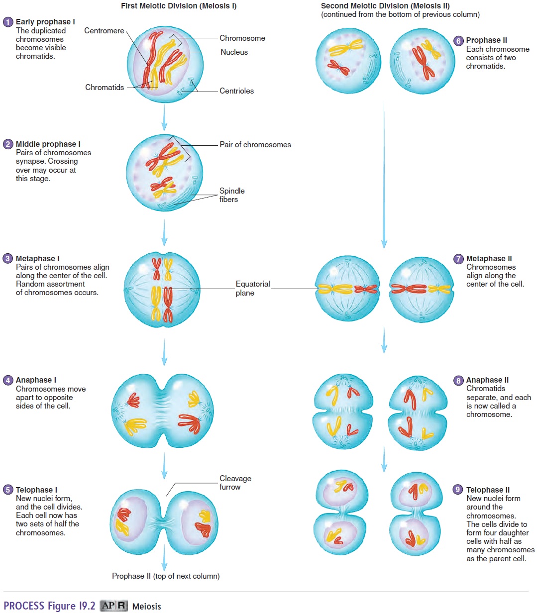
The chromosomes align along the center of the cell (figure 19.2, step 3), and then the pairs of chromosomes are separated to each side of the cell (figure 19.2, step 4). As a consequence, when meiosis I is complete, each daughter cell has 1 chromosome from each of the pairs (figure 19.2, step 5), or 23 chromosomes. Each of the 23 chromosomes in each daughter cell consists of 2 chromatids joined by a centromere.
It is during the first meiotic division that the chromosome number is reduced from 46 (23 pairs) to 23 chromosomes. The first meiotic division is therefore called a reduction division.
The second meiotic division is similar to mitosis. The chro-mosomes, each consisting of 2 chromatids (figure 19.2, steps 6 and 7), align along the center of the cell. Then the chromatids separate at the centromere, and each daughter cell receives 1 of the chromatids from each chromosome (figure 19.2, steps 8 and 9). When the centromere separates, each of the chromatids is called a chromosome. Consequently, each of the 4 daughter cells produced by meiosis contains 23 chromosomes.
During fertilization, the zygote receives 1 set of chromosomes (23) from each parent. Although half the genetic material of a zygote comes from each parent, the genetic makeup of each zygote is unique.
Related Topics