Chapter: Ophthalmology: Vitreous Body
Vitreous Body
Vitreous Body
Basic Knowledge
Importance of the vitreous body for the eye:
The vitreous body stabilizesthe globe although
the eye can remain intact without the vitreous body (see vitrectomy). It also
prevents retinal detachment.
Embryology:
The development of the vitreous body can be divided into
threephases:
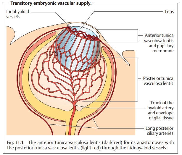
❖ First phase (first month of pregnancy; fetus measures 5 – 13 mm craniumto
coccyx): The primary vitreous forms
during this period. This phase is characterized by the entry of mesenchyme into
the optic cup through the embryonic choroidal fissure. The main function of the primary vitreous is to
supply the developing lens with nourishment. In keeping with this func-tion, it
consists mainly of a vascular plexus, the anterior
and posteriortunica vasculosa lentis, that covers the anterior and
posterior surfaces ofthe lens. This vascular plexus is supplied by the hyaloid
artery and its branches (Fig. 11.1).
This vascular system and the primary vitreous regress as the posterior lens
capsule develops at the end of the second month of pregnancy.
❖ Second phase (second month of pregnancy; fetus measures 14 – 70 mmcranium to
coccyx): The secondary vitreous
forms during this period. This avascular vitreous body consisting of fine
undulating collagen fibers develops from what later becomes the retina. In
normal development it expands to compress the central primary vitreous into a
residual central canal (hyaloid canal
or Cloquet’s canal).
❖ Third phase (third month of pregnancy; fetus measures 71 – 110 mmcranium to
coccyx): The tertiary vitreous
develops from existing struc-tures in the secondary vitreous. The secondary
vitreous remains. The zonule fibers that
form the suspensory ligament of the lens develop duringthis period.
Composition of the vitreous body:
The gelatinous vitreous body consists of98% water and 2% collagen and hyaluronic acid. It fills the vitreous chamber, which accounts for approximately two-thirds of the total volume of the eye.
Stabilization and confines of the vitreous body:
With their high negativeelectrostatic
potential, the hyaluronic acid molecules fill the three-dimen-sional collagen
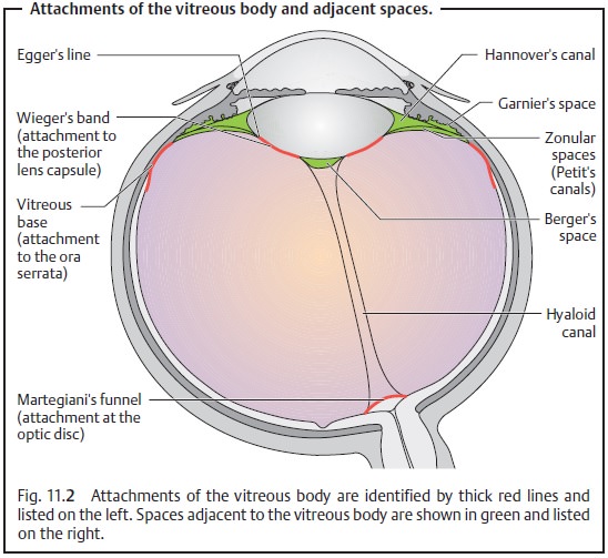
fiber network and provide mechanical stability. Condensationof peripheral collagen fibrils creates aboundary membrane(hyaloid mem-brane), which isnot
a basement membrane.It is attached to adjacent struc-tures at the following
locations (Fig. 11.2):
❖At the ligament of Wieger along the posterior capsule of the lens.
❖At the vitreous base at the ora serrata.
❖ At the
funnel of Martegiani (approximately 10 µm wide) surrounding
the periphery of the optic disk.
The connections between the vitreous body and retina are generally loose although there may be firm focal adhesions. These firmer focal attachments cause problems during vitreous detachment because they do not permit the vitreous body to become completely detached. The focal adhesions between the vitreous body and retina produce focal traction forces that act on the retina and can cause retinal tears and detachment.
Neurovascular supply:
The vitreous body contains neither blood vessels nornerves. As a
result, pathogens can multiply undisturbed for a relatively long time before
the onset of an immune response from adjacent structures.
Related Topics