Chapter: Ophthalmology: Vitreous Body
Vitreous Body: Aging Changes

Aging Changes
Synchysis
The regular arrangement of collagen fibers
gradually deteriorates in middle age. The fibers condense to flattened
filamentous structures. This process, known as liquefaction, creates small
fluid-filled lacunae in the central vit-reous body that initially are largely
asymptomatic (patients may report floaters). However, once liquefaction has
progressed beyond a certain point, the vitreous body can collapse and detach
from the retina.
Vitreous Detachment
Definition
Complete or partial detachment of the vitreous
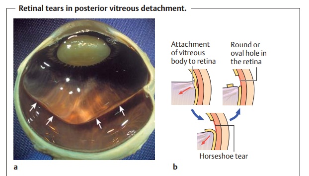
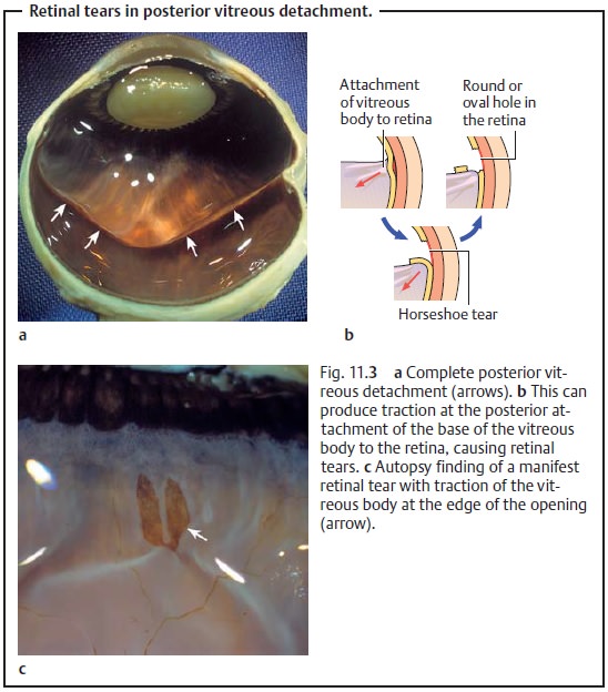
body from its underlying tissue. The most common form is posterior vitreous detachment (see Fig. 11.3a); anterior or basal vitreous detachment is much rarer.

Epidemiology:
Six percent of patients between the ages of 54 and 65 and 65%of
all patients between the ages of 65 and 85 have posterior vitreous detach-ment.
Patients with axial myopia have a predisposition to early vitreous detachment.
Presumably the vitreous body collapses earlier in these patients because it
must fill a “longer” eye with a larger volume.
Etiology:
Liquefaction causes collapse of the vitreous body. This
usuallybegins posteriorly where the attachments to the underlying tissue are
least well developed. Detachment in the anterior region (anterior vitreous detach-ment) or in the region of the vitreous
base (basal vitreous detachment)
usuallyonly occurs where strong forces act on the globe as in ocular trauma.
Symptoms and findings:
Collapse of the vitreous bodyleads to vitreous densi-ties that the patient
perceives as mobile opacities. These floaters (also known as flies or cobwebs)
may take the form of circular or serpentine lines or points. The vitreous body
may detach partially or completely from the retina. An increased risk of
retinal detachment is present only with partial
vitreousdetachment. In this case, the vitreous body and retina remain
attached, withthe result that eye movements in this region will place traction
on the retina. The patient perceives this phenomenon as flashes of light. If the traction on the retina becomes too strong,
it can tear (see retinal tears in posterior vit-reous detachment, Fig. 11.3b – c). This increases the risk of
retinal detachment and vitreous bleeding from injured vessels.
Floaters and especially flashes of light require thorough
examination of the ocular fundus to exclude a retinal tear.
Diagnostic considerations:
The symptoms of vitreous detachment requireexamination of the
entire fundus of the eye to exclude a retinal defect. In cases such as lens
opacification or vitreous hemorrhage where visualization is not possible, an
ultrasound examination is required to evaluate the vitreous body and retina.
Vitreous detachment in the region of the attachment at the optic
disk (funnel of Martegiani) will
appear as a smokey ring (Weiss’ ring)
under ophthalmoscopy.
Treatment:
The symptoms of vitreous detachment resolve spontaneouslyonce
the vitreous body is completely detached. However, the complications that can
accompany partial vitreous detachment require treatment. These include retinal
tears, retinal detachment (for treatment see Ret-ina), and vitreous hemorrhage.
Related Topics