Chapter: Ophthalmology: Uveal Tract (Vascular pigmented layer)
Uveal Tract (Vascular pigmented layer): Basic Knowledge
Uveal Tract (Vascular
pigmented layer)
Basic Knowledge
Structure: The uveal tract (also known as the vascular pigmented
layer,vascular tunic, and uvea) takes its name from the Latin uva (grape) because the dark
pigmentation and shape of the structure are reminiscent of a grape. The uveal
tract consists of the following structures:
❖ Iris,
❖ Ciliary body,
❖ Choroid.
Position: The uveal tract lies between the sclera and retina.
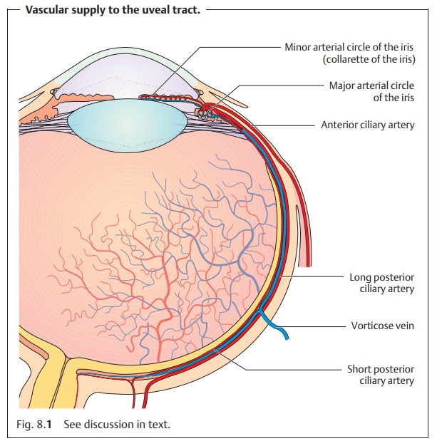
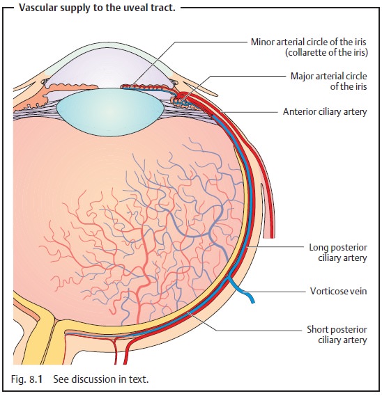
Neurovascular supply: Arterial supplyto the uveal tract is provided by theophthalmic artery.
❖Theshort
posterior ciliary arteries enter the eyeball with the optic nerve and
supply the choroid.
❖ Thelong posterior ciliary arteries course
along the interior surface of the sclera to the ciliary body and the iris.
They form the major arterial circle at the root of the iris and the minor
arterial circle in the collarette of the iris.
The anterior
ciliary arteries originate from the vessels of the rectus muscles and
communicate with the posterior ciliary
vessels.
Venous blood drains through four to eightvorticose
or vortex veinsthatpenetrate the sclera posterior to the equator and join
the superior and inferior ophthalmic veins (Fig. 8.1). Sensory supply is
provided by the long and shortciliary
nerves.
Iris
Structure and function: The iris consists of two layers:
❖ Theanterior mesodermal stromal layer.
❖ Theposterior ectodermal pigmented epithelial layer.
The posterior layer is opaque and protects the eye against excessive incident light. The anterior surface of the lens and the pigmented layer are so close together near the pupil that they can easily form adhesions in inflammation.
The collarette of the iris covering the minor arterial circle of the
iris divides the stroma into pupillary
and ciliary portions. The pupillary
portion contains the sphincter muscle, which is supplied by parasympathetic nerve fibers, and the dilator pupillae muscle, supplied by sympathetic nerve fibers. These muscles regulate
the contraction and dilation of the pupil so that the iris may be regarded as
the aperture of the optical system of the eye.
Pupil dilation is sometimes sluggish in
preterm infants and the newborn because the dilator pupillae muscle develops
relatively late.
Surface: The normal iris has a richly textured surface
structure withcrypts(tissue gaps)
and interlinked trabeculae. A faded
surface structure can be a sign of
inflammation (see iridocyclitis).
Color: The color of the iris varies in the individual according to themelanincontent of the melanocytes (pigment
cells) in thestromaandepithelial layer.Eyes with a high
melanin content are dark brown, whereas eyes with less melanin are
grayish-blue. Caucasians at birth
always have a grayish-blue iris as the pigmented
layer only develops gradually during the first year of life. Even in albinos (see impaired melanin
synthesis), the eyes have a grayish-blue iris because of the melanin
deficiency. Under slit lamp retroillumination they appear reddish due to the
fundus reflex.
Ciliary Body
Position and structure: Theciliary bodyextends
from the root of the iris tothe ora serrata, where it joins the choroid. It
consists of anterior pars plicata and
the posterior pars plana, which lies
3.5 mm posterior to the limbus. Numerous ciliary
processes extend into the posterior chamber of the eye. The suspensory
ligament, the zonule, extends from the pars plana and the inter-vals between
the ciliary processes to the lens capsule.
Function: Theciliary muscleis
responsible foraccommodation.The
double-layered epithelium covering the
ciliary bodyproduces the aqueous humor.
Choroid
Position and structure: The choroid is themiddle
tunic of the eyeball. It isbounded on the interior by Bruch’s membrane. The choroid is highly vascu-larized, containing a
vessel layer with large blood vessels and a capillary layer. The blood flow
through the choroid is the highest in the
entire body.
Function: The choroidregulates
temperatureand suppliesnourishment
tothe outer layers of the retina.
Related Topics