Chapter: Ophthalmology: Uveal Tract (Vascular pigmented layer)
Choroiditis

Choroiditis
Epidemiology:
There are few epidemiologic studies of choroiditis.
Theannual incidence is assumed to be four cases per 100 000 people.
Etiology:
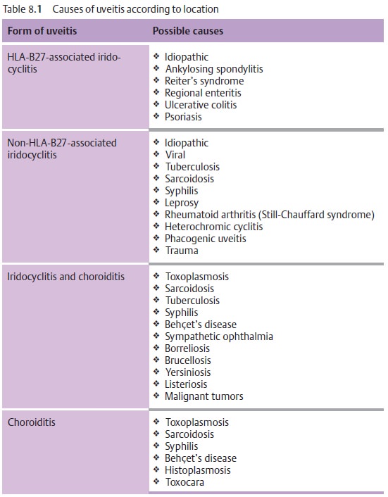
See Table 8.1.

Symptoms:
Patients are free of pain, although they
report blurred vision andfloaters.
Choroiditis is painless as the choroid is
devoid of sensory nerve fibers.
Diagnostic considerations:
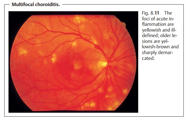
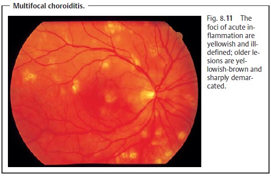
Ophthalmoscopy reveals isolated or multiplechoroiditis foci. In acute disease they appear as ill-defined white dots (Fig. 8.11). Once scarring has occurred the foci are sharply demarcated with a yellowish-brown color. Occasionally the major choroidal vessels will be vis-ible through the atrophic scars.

No cells will be found in the vitreous body in aprimary choroidal
process.However,
inflammation proceeding from the retina (retinochoroiditis) will exhibit cellular infiltration of the vitreous body.
Differential diagnosis:
This disorder should be distinguished from
retinalinflammations, which are accompanied by cellular infiltration of the
vitreous body and are most frequently caused by viruses or Toxoplasma gondii.
Treatment:
Choroiditis is treated either with antibiotics
or steroids, depend-ing on its etiology.
Prognosis:
The inflammatory foci will heal within two to
six weeks and formchorioretinal scars. The scars will result in localized
scotomas that will reduce visual acuity if the macula is affected.
Related Topics