Chapter: Ophthalmology: Uveal Tract (Vascular pigmented layer)
Uveal Tract (Vascular pigmented layer): Examination Methods
Examination Methods
The slit
lamp is used to examine the surface of the iris under a focused beam of light. Normally no vessels will be visible.
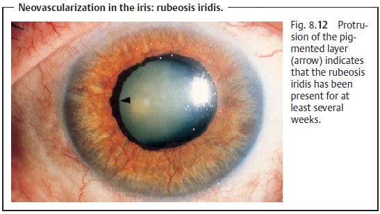
Iris vessels are only visible in atrophy of
the iris, inflammation, or as neovascularization in rubeosis iridis (see Fig.
8.12).

Where vessels are present, they can be
visualized by iris angiography after
intravenous injection of fluorescein sodium dye.
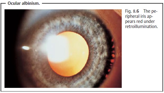
Defects in the pigmented layer of the iris appear red under retroillumina-tion with a
slit lamp (see Fig. 8.6). Slit lamp biomicroscopy visualizes
indi-vidual cells such as melanin cells at 40-power magnification.

The anterior chamber
is normally transparent. Inflammation
can increase the permeability of the vessels of the iris and compromise the barrier
between blood and aqueous humor. Opacification of the aqueous humor by
proteins may be observed with the aid of a slit
lamp when the eye is illumi-nated with a lateral focal beam of light
(Tyndall effect). This method can also be used to diagnose cells in the anterior chamber in the presence of inflamma-tion.
Direct inspection of the root of the iris is not possible because it does not lie within the line of
sight. However, it can be indirectly visualized by gonios-copy. Inspection of theposterior portion of the
pars planarequires a three-mirror lens. The globe is also
indented with a metal rod to permit visualizationof this part of the ciliary
body (for example in the presence of a suspected malignant melanoma of the
ciliary body).
The pigmented epithelium of the retina permits
only limited evaluation of the choroid by ophthalmoscopy and
fluorescein angiography or
indocyanine green angiography. Changes in the choroid such as tumors or
hemangiomas can be visualized by ultrasound examination. Where a tumor is suspected, transillumination of the eye is indicated. After administration
of topical anes-thesia, a fiberoptic light source is placed on the eyeball to visualize the shadowof the tumor on the red
of the fundus.
Related Topics