Chapter: Clinical Dermatology: The function and structure of the skin
The dermo-epidermal junction

The dermo-epidermal
junction
The
basement membrane lies at the interface between the epidermis and dermis. With
light microscopy it can be highlighted using a periodic acid–Schiff (PAS)
stain, because of its abundance of neutral mucopolysac-charides. Electron microscopy
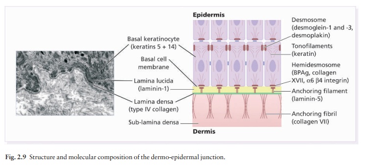
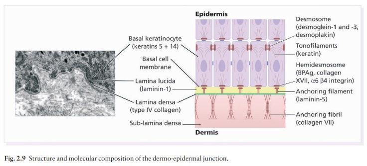
(Fig. 2.9) shows that the lamina densa (rich in type IV collagen) is
separatedfrom the basal cells by an electron-lucent area, the lamina
lucida. The plasma membrane of basal cellshas hemidesmosomes
(containing bullous pemphigoid antigens, collagen XVII and α6 β4
integrin). The lamina lucida contains the adhesive macromolecules, laminin-1,
laminin-5 and entactin. Fine anchoringfilaments (of
laminin-5) cross the lamina lucida andconnect the lamina densa to the plasma
membrane of the basal cells. Anchoring fibrils (of type
VII collagen), dermal microfibril bundles and single small collagen fibres
(types I and III), extend from the papillary dermis to the deep part of the
lamina densa.

Laminins,
large non-collagen glycoproteins pro-duced by keratinocytes, aided by entactin,
promote adhesion between the basal cells above the lamina lucida and type IV
collagen, the main constituent of the lamina densa, below it. The laminins act
as a glue, helping to hold the epidermis onto the dermis. Bullous pemphigoid
antigens (of molecular weights 230 and kDa) are synthesized by basal cells and
are found in close association with the hemidesmosomes and laminin. Their
function is unknown but antibodies to them are found in pemphigoid, a
subcutan-eous blistering condition.
The
structures within the dermo-epidermal junction provide mechanical support,
encouraging the adhesion, growth, differentiation and migration of the
overlying basal cells, and also act as a semipermeable filter that regulates
the transfer of nutrients and cells from dermis to epidermis.
Related Topics