Chapter: Human Neuroanatomy(Fundamental and Clinical): Internal Capsule and Commissures of the Brain
The Lateral Geniculate Body
The Lateral Geniculate Body
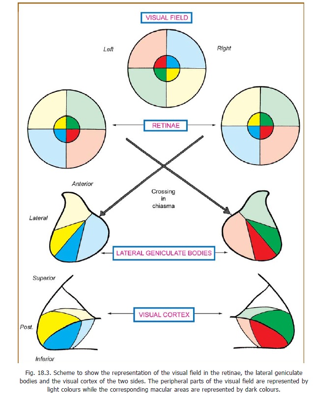
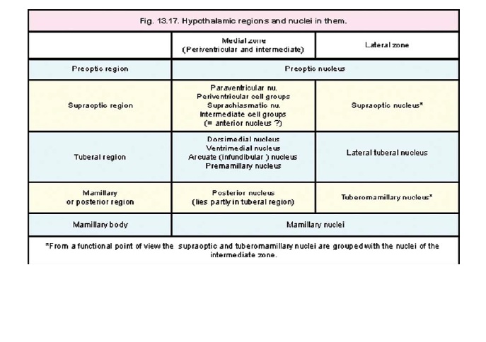
Some aspects of the structure of the lateral geniculate body have been considered. It has been seen that the grey matter of this body is split into six laminae (Fig. 13.17). Fibres from the eye of the same side end in laminae 2, 3, and 5; while those from the opposite eye end in laminae 1, 4, and 6. The macular fibres end in the central and posterior part of the lateral geniculate body, and this area is relatively large (Fig. 18.3). Fibres from the peripheral parts of the retina end in the anterior part of the lateral geniculate body. The upper half of the retina is represented laterally, and the lower half of the retina is represented medially. Specific points on the retina project to specific points in the lateral geniculate body. In turn, specific points of this body project to specific points in the visual cortex. In this way a point to point relationship is maintained between the retinae and the visual cortex.
Recent studies of synaptic patterns within the lateral geniculate body indicate that this nucleus is not to be regarded as a simple relay station and that various influences may modify the conduction of impulses through it. In this connection it may be noted that the lateral geniculate body receives afferents from the visual cortex.
Related Topics