Chapter: Human Neuroanatomy(Fundamental and Clinical): Internal Capsule and Commissures of the Brain
Optic Nerve, Optic Chiasma & Optic Tract
Optic Nerve, Optic Chiasma & Optic Tract
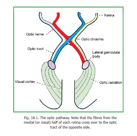
The optic nerve is made up of axons of the ganglion cells of the retina. These axons are at first unmyelinated. The fibres from all parts of the retina converge on the optic disc. In this region the sclera has numerous small apertures and is, therefore, called the lamina cribrosa (crib = sieve). Bundles of optic nerve fibres pass through these apertures. Each fibre acquires a myelin sheath as soon as it pierces the sclera. The fibres of the nerve arising from the four quadrants of the retina maintain the same relative position within the nerve. The fibres arising from the macula are numerous and form the papillomacular bundle. Close to the eyeball, the macular fibres occupy the lateral part of the nerve, but by the time they reach the chiasma they lie in the central part of the nerve.

The fibres of the optic nerve arising in the nasal half of each retina enter the optic tract of the opposite side after crossing in the chiasma. Fibres from the temporal half of each retina enter the optic tract of the same side (Fig. 18.1). Thus the right optic tract comes to contain fibres from the right halves of both retinae, and the left tract from the left halves. In other words, all optic nerve fibres carrying impulses relating to the left half of the field of vision are brought together in the right optic tract and vice versa. We have already noted that each optic tract carries these fibres to the lateral geniculate body of the corresponding side.
Related Topics