Chapter: Genetics and Molecular Biology: Biological Assembly, Ribosomes and Lambda Phage
Structure of the Lambda Particle
The Structure of the Lambda Particle
The physical structure of lambda phage and the
organization of the genetic map of lambda show the same relationship as the
physical structure of the ribosome and the organization of the genetic map of
ribosomal proteins. The genes for proteins that lie near one another in the
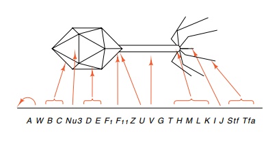
particles often lie near one another in the genetic map (Fig. 21.15). Even
though a single species of a polycistronic messenger is synthesized that
includes all the phage late genes, they are translated at vastly different
efficiencies closely paralleling the numbers of the different types of protein
molecules that are found in the mature phage particle.
The A protein, often denoted pA, in conjunction
with pNu1 is respon-sible for producing the cohesive ends of the DNA. Proteins
pD and pE are the major capsid proteins. Protein pE plus Nu3 followed by pB and
pC is able to form a precursor of the head, pNu3 is then degraded and pD is
added. In the final structure, protein pD is interspersed with equal numbers of
protein pE, that is, pD and pE are the two major capsid proteins. Protein pFII as well
as protein pW are involved with steps preparing the head assembly for receipt
of the tail. pFII provides a specificity for the type of tail that is attached to the
head. Protein pV is the major structural protein of the tail. Proteins pH, pM,
pL, and pK are

Figure
21.15 The genetic map ofsome genes
involved with forming the head and tail of lambda and the locations of the gene
products in the physical structure of the head and tail.
Related Topics