Chapter: 11th Zoology : Chapter 7 : Body Fluids and Circulation
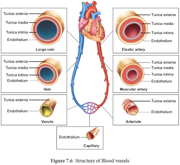
Structure of blood vessels
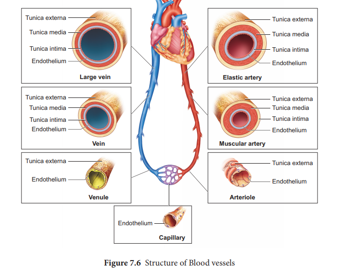
Structure of
blood vessels
The
vessels carrying the blood are of three types; they are the arteries, veins and
capillaries. These vessels are hollow structures and have complex walls
surrounding the lumen. The blood vessels in humans are composed of three
layers, tunica intima, tunica media and tunica externa. The inner layer, tunica intima or tunica interna supports the vascular endothelium, the
middle layer, tunica media is composed of smooth muscles and an extra cellular
matrix which contains a protein, elastin. The contraction
and relaxation of the smooth muscles results in vasoconstriction and
vasodilation. The outer layer, tunica externa or tunica adventitia is composed
of collagen fibres. The structure of blood vessels is illustrated in Figure
7.6.
Arteries
The blood
vessels that carry blood away from the heart are called arteries. The arteries
usually lie deep inside the body. The walls of the arteries are thick, non
collapsible to withstand high pressure. Valves are absent and have a narrow
lumen. All arteries carry oxygenated blood, except the pulmonary artery. The
largest artery, the aorta (2.5 cm in diameter and 2 mm thick) branch into
smaller arteries and culminates into the tissues as feed arteries. In the
tissues the arteries branches into arterioles.
As blood
enters an arteriole it may have a pressure of 85 mm Hg (11.3 KPa) but as it
leaves and flows into the capillary, the pressure drops to 35 mm Hg (4.7 KPa).
(Note 1 mm Hg =0.13 KPa. SI unit of Hg
is KiloPascal (KPa)). Arterioles are small, narrow, and thin walled which are
connected to the capillaries. A small sphincter lies at the junction between
the arterioles and capillaries to regulate the blood supply. Arteries do not
always branch into arterioles, they can also form anastomoses.
Capillaries
Capillary
beds are made up of fine networks of capillaries. The capillaries are thin
walled and consist of single layer of
Tunica media and elastin fibres are absent. The capillary beds are
the site for exchange of materials between blood and tissues. The walls of the
capillaries are guarded by semilunar valves. The blood volume in the
capillaries is high but the flow of blood is slow. Mixed blood (oxygenated and
deoxygenated) is present in the capillaries. The capillary bed may be flooded
with blood or may be completely bypassed depending on the body conditions in a
particular organ.
Veins
Veins
have thinner walls and a larger lumen and hence can be easily stretched. They
carry deoxygenated blood except, the pulmonary vein. The blood pressure is low
and the lumen has a wide wall which is collapsible. Tunica media is thinner in
veins than in arteries. Unidirectional flow of blood in veins is due to the
presence of semilunar valves that prevents backflow of blood. Blood samples are
usually taken from the veins rather than artery because of low pressure in the
veins.
1. Coronary blood vessels
Blood
vessels that supply blood to the cardiac muscles with all nutrients and removes
wastes are the coronary arteries and veins. Heart muscle is supplied by two
arteries namely right and left coronary arteries. These arteries are the first
branch of the aorta. Arteries usually surround the heart in the manner of a
crown, hence called coronary artery (L. Corona
- crown).
Right
ventricle and posterior portion of left ventricle are supplied by the right
coronary artery. Anterior and lateral part of the left ventricle is supplied by
the left coronary arteries.
Related Topics