Zoology - Body Fluids and Circulation: Important Questions | 11th Zoology : Chapter 7 : Body Fluids and Circulation
Chapter: 11th Zoology : Chapter 7 : Body Fluids and Circulation
Body Fluids and Circulation: Important Questions
Body
Fluids and Circulation
Evaluation
1. What is the function of lymph?
a.
Transport of O2 into brain
b.
Transport of CO2 into lungs
c. Bring interstitial fluid in
blood
d.
Bring RBC and WBC in lymph node
2. Which one of the following
plasma proteins is involved in the coagulation of blood?
a.
Globulin
b. Fibrinogen
c.
Albumin
d.
Serum amylase
3. Which of the following WBCs are
found in more numbers?
a.
Eosinophil
b. Neutrophil
c.
Basophil
d.
Monocyte
4. Which of the following is not
involved in blood clotting?
a.
Fibrin
b.
Calcium
c.
Platelets
d. Bilirubin
5. Lymph is colourless because
a.
WBC are absent
b.
WBC are present
c.
Heamoglobin is absent
d. RBC are absent
6. Blood group is due to the
presence or absence of surface
a.
Antigens on the surface of WBC
b.
Antibodies on the surface of RBC
c. Antigens of the surface of RBC
d.
Antibodies on the surface of WBC
7. A person having both antigen A
and antigen B on the surface of RBCs belongs to blood group
a.
A
b.
B
c. AB
d.
O
8. Erythroblastosis foetalis is due
to the destruction of
a. Foetal RBCs
b.
Foetus suffers from atherosclerosis
c.
Foetal WBCs
d.
Foetus suffers from mianmata
9. Dub sound of heart is caused by
a.
Closure of atrio-ventricular valves
b.
Opening of semi-lunar valves
c. Closure of semi-lunar values
d.
Opening of atrio-ventricular valves.
10. Why is the velocity of blood
flow the lowest in the capillaries?
a.
The systemic capillaries are supplied by the left ventricle, which has a lower
cardiac output than the right ventricle.
b.
Capillaries are far from the heart, and blood flow slows as distance from the
heart increases.
c. The total surface area of the
capillaries is larger than the total surface area of the arterioles.
d.
The capillary walls are not thin enough to allow oxygen to exchange with the
cells.
e.
The diastolic blood pressure is too low to deliver blood to the capillaries at
a high flow rate.
11. An unconscious patient is
rushed into the emergency room and needs a fast blood transfusion. Because
there is no time to check her medical history or determine her blood type,
which type of blood should you as her doctor, give her?
a.
A-
b.
AB
c. O+
d.
O-
12. Which of these functions could
or could not be carried out by a red blood cell? Briefly justify your answer.
a.
Protein synthesis
b.
Cell division
c.
Lipid synthesis
d.
Active transport
13. At the venous end of the
capillary bed, the osmotic pressure is
a. Greater than the hydrostatic
pressure
b.
Result in net outflow of fluids
c.
Results in net absorption of fluids
d.
No change occurs.
14. A patient’s chart reveals that
he has a cardiac output of 7500mL per minute and a stroke volume of 50 mL. What
is his pulse rate (in beats / min)
a.
50
b.
100
c. 150
d.
400
15. At any given time there is more
blood in the venous system than that of the arterial system. Which of the
following features of the veins allows this?
a.
relative lack of smooth muscles
b.
presence of valves
c. proximity of the veins to
lymphatic’s
d.
thin endothelial lining
16.
Distinguish between arteries and veins
17.
Distinguish between open and closed circulation
18.
Distinguish between mitral valve and semi lunar valve
19.
Right ventricular wall is thinner than the left ventricular wall. Why?
20.
What might be the effect on a person whose diet has less iron content?
21.
Describe the mechanism by which the human heart beat is initiated and
controlled.
22.
What is lymph? Write its function.
23.
What are the heart sounds? When and how are these sounds produced?
24.
Select the correct biological term.
Lymphocytes,
red cells, leucocytes, plasma, erythrocytes, white cells, haemoglobin,
phagocyte, platelets, blood clot.
a.
Disc shaped cells which are concave on both sides - erythrocytes
b.
Most of these have a large, bilobed nucleus - Lymphocytes
c.
Enable red cells to transport blood
d.
The liquid part of the blood - plasma
e.
Most of them move and change shape like an amoeba. - phagocyte
f.
Consists of water and important dissolved substances. - plasma
g.
Destroyed in the liver and spleen after circulating in the blood for four
months. - erythrocytes
h.
The substances which gives red cells their colour. - haemoglobin
i.
Another name for red blood cells. - erythrocytes
j.
Blood that has been changed to a jelly. - blood
clot
k.
A word that means cell eater. - phagocyte
l.
Cells without nucleus. - erythrocytes /phagocyte
m.
White cells made in the lymphatic tissue. - Lymphocytes
n.
Blocks wound and prevent excessive bleeding. - blood clot
o.
Fragment of cells which are made in the bone marrow. - red cells
p.
Another name for white blood cells. - leucocytes
q.
Slowly releases oxygen to blood cells. - haemoglobin
r.
Their function is to help blood clot in wounds. - platelets
25.
Select the correct biological term.
Cardiac
muscle, atria, tricuspid systole, auricles, arteries, diastole, ventricles,
bicuspid valve, pulmonary artery, cardiac cycle, semi lunar valve, veins,
pulmonary vein, capillaries, vena cava, aorta.
a.
The main artery of the blood. - aorta
b.
Valves between the left atrium and ventricle. - bicuspid valve
c.
Technical name for relaxation of the heart. - diastole
d.
Another name for atria. - auricles
e.
The main vein. - vena cava
f.
Vessels which carry blood away from the heart. - arteries
g.
Two names for the upper chambers of the heart. – atria/ auricles
h.
Thick walled chambers of the heart. - ventricles
i.
Carries blood from the heart to the lungs. - pulmonary artery
j.
Takes about 0.8 sec to complete. - cardiac
cycle
k.
Valves situated at the point where blood flows out of the heart. - semi lunar valve
l.
Vessels which carry blood towards the heart. - veins
m.
Carries blood from the lungs to the heart. - pulmonary vein
n.
The two lower chambers of the heart. - ventricles
o.
Prevent blood from re entering the ventricles after entering the aorta. . - semi lunar valve
p.
Technical name for one heart beat. - Cardiac
muscle
q.
Valves between right atrium and ventricles. - tricuspid systole
r.
Technical name for contraction of the heart. - Systole
s.
Very narrow blood vessels. - Capillaries
26.
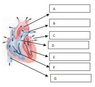
Name and Label the given diagrams to show A, B, C, D, E, F, and G

Glossary
Blood vessels
serve as a passage way through which the blood is directed and distributed from
the heart to all parts of the body and subsequently returned to the heart.
Pulmonary circulation
– consists of closed loop of vessels carrying blood between the heart and
lungs.
Systemic circulation
– is a circuit of vessels carrying blood between the heart and other parts of
body systems.
Cardio pulmonary
resuscitation (CPR) – Serves as a life saving measure until
appropriate therapy can restore the heart to normal function.
Aorta
– A single large artery carrying blood away from the left ventricle.
Bicuspid valve
– also called mitral valve. Left Auricular ventricular valve with two flaps
that is present between the left auricle and left ventricle.
Tricuspid valve
– right auricular valve with three flaps that is present between the right
auricle and right ventricle.
Chordate tendineae
– these are chords that extend from the edge of each flap and attach to the
papillary muscles that prevent the AV valves from being forced to open due to
high ventricular pressure.
Papillary muscles
– small nipple shaped muscles protrude from the inner surface of the
ventricular walls. Papilla means ‘nipple’.
Sinoatrial node (SA
node), – a small, specialised region in the right atrial
wall near the opening of the superior vena cava
Atrioventricular node
(AV node), – a small bundle of specialized cardiac muscle
cells locted at the base of the right atrium near the septum, just above the
junction of the atria and ventricles.
Bundle of His
– (atrioventicular bundle), a tract of specialized cells that originates at the
AV node and enters the interventricular septum
Purkinje fibres
– small terminal fibres that extend from the bundle of His and spread
throughout the ventricular myocardium
Stroke volume (SV)
– The amount of blood pumped out of each ventricle with each contraction, SV =
EDV-ESV
Isovolumetric
ventricular contraction – Isovolumetric means constant
volume and length. During ventricular contraction, when all valves are closed,
no blood can enter or leave the ventricle during this time. Because no blood
leaves or enters the ventricles the ventricular chamber has a constant volume
and the muscle fibres stay at a constant length.
End systolic volume
(ESV) – The ventricles do not empty completely during
ejection, only half of the blood within the ventricle at the end of diastole is
pumped out during subsequent systole. The amount of blood left in the ventricle
at the end of systole when ejection is complete is called ESV.
End diastolic volume
(EDV) – The volume of blood in the ventricle at the end
of diastole is known as the end diastolic volume.
Lub sound
– is associated with the closure of the AV valves.
Dub sound
– is associated with the closure of the semilunar valves.
Chordae tendinae
– tendon like cords which are connected to the tip of the cuspid valves
Diastole
– Relaxation of heart chambers
Endocardium
– Inner cardiac muscle
Epicardium
– outer cardiac muscle
Inter ventricular
septum – Partition between right and left ventricle
Interatrial septum
– Partition between right and left atria
Left atrioventricular
valve – Bicuspid valve or Mitral valve
Related Topics