Chapter: 11th Microbiology : Chapter 3 : Stains and Staining Methods
Special Staining - Endospore Staining
Special Staining – Endospore
Staining
Endospores are highly resistant
structures produced by some bacteria during unfavourable environment
conditions. Endospore formation is a distinguishing feature of aerobic genera Bacillus and anaerobic genera Clostridium. The size, shape and
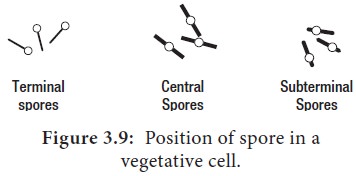
position of the spore (Figure 3.9) are relatively constant characteristics of a
given species and are important in identifying the species within genera. The
position of spore in the cell may be terminal, central or sub-terminal. Figure
3.9 shows the position of spores in a vegetative cell.

Endospores cannot be stained by
ordinary methods, such as simple staining and Gram staining, because the dyes
do not penetrate the wall of the endospore. If simple stains are used, the
vegetative body of the bacillus is deeply coloured, whereas the spore is
unstained and appears as a clear area in the organism.
By vigorous staining procedure, the
dye can be introduced into the spore. Once stained, the spore tends to retain
the dye even after treatment with decolorizing agents. The most commonly used
endospore staining procedure is the Schaeffer Fulton endospore staining method.
Malachite green, the primary stain, is applied to a heat fixed smear and heated
to steaming for about 5 minutes. Heat helps the stain to penetrate the
endospore wall. Then the preparation is washed for about 30 seconds with water.
Next safranin, a counterstain is applied to the smear to stain the portions of
the cell other than endospores.
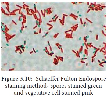
In a properly prepared smear, the
endospores appear green within red cells (Figure 3.10). Endospores are highly
refractive. They can be detected under the light microscope when unstained, but
cannot be differentiated from inclusions of stored material without a special
stain.


Related Topics