Movements in Animals | Chapter 19 | 8th Science - Skeleton System | 8th Science : Chapter 19 : Movements in Animals
Chapter: 8th Science : Chapter 19 : Movements in Animals
Skeleton System
Skeleton System
The skeleton system provides the
hard structure or framework to the human body which supports and protects the
body. It is composed of connective tissues like bones, cartilage, tendons and
ligaments. If the skeleton is without joints, no movement would take place and
the significance of human body will be no more than a stone. On the basis of
presence in the body, skeleton is of two types.
Exoskeleton
It is the skeleton that is found on
the exterior layer of the body and it basically originates from embryonic
ectoderm or mesoderm. Like scales in the fishes, outer hard layer of the
tortoise and feathers of the birds it protects and preserves the inner organs.
Endoskeleton
It is the skeleton that is found
inside the human body and it originates from the mesoderm. These are found in
almost all vertebrates and form the main body structure.
1. Functions of skeleton
The skeletal system serves five
important functions in the human body.
1. It provides structure and shape
to the body.
2. It supports and surrounds the
internal organs of the body.
3. Calcium and phosphorus, the two
minerals that the body needs for important regulatory functions, are stored
inside the bones.
4. Red blood cells are produced in
the bone marrow.
5. The bones of the skeletal system
act as levers for muscular action. Muscular movement would not be possible
without tendons (fibrous cords of
tissue that attach muscle to bone) and ligaments
(fibrous cords of tissue that attach bone to bone).
• The femur or
thighbone is the longest and strongest bone of the human skeleton. • The stapes
in the middle ear is the smallest and lightest bone of the human skeleton.
2. Constituents of
skeleton
Human skeleton consists of bone,
cartilages and ligaments. Bones comprise the hard framework of the body.
Cartilages are the supporting and connecting structures. For example, the
cartilage supports the projecting external ears and the tip of the nose.
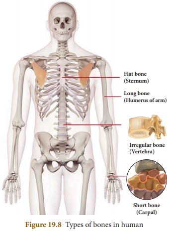
Ligaments bind the bones together. There are different types of bones in human
skeletal system. They are:
Long
bones: Found in arms and
legs.
Short
bones: Found in wrist
ankle, vertebral column.
Flat
bones: Found in skull,
ribs, shoulder and hips.
Irregular
bones: Found in spine and
vertebral column, mandible, palatine, inferior nasal concha, and hyoid.
3. Parts of skeleton
The skeletal system is composed of
bones and the related structures that aid body movement. It is divided into two
major parts: the axial skeleton and the appendicular skeleton.
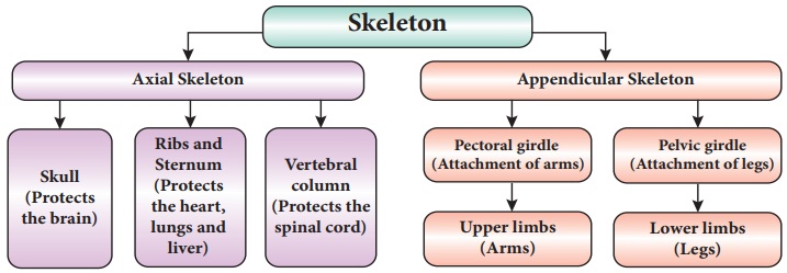
I. Axial skeleton
The axial skeleton consists of the bones along the axis, or central
line of the human body. The axial skeleton consists of the skull, facial bones,
sternum, ribs, and vertebral column.
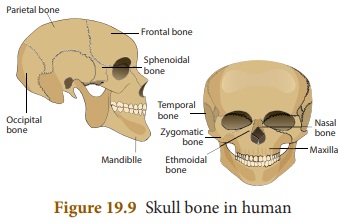
a.
Skull
Skull is a hard structure made up of
small bones. It is formed by 22 bones out of which 8 bones are fixed together
to form the cranium and 14 bones fuse to form the face. The only bone which has
movable joint is the lower jaw. This movable joint is supported by muscles and
ligaments. Skull placed on the top of the backbone can be moved up, down and
sidewards.
b.
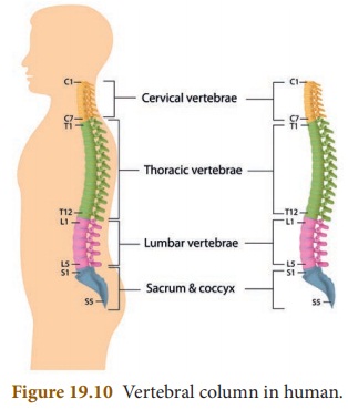
Vertebral column
Vertebral column running at the back
of the body is also called as spine or the backbone. It is in the trunk region
to offer support to the upper part of the body. Vertebral column is made up of
individual bones called as vertebrae. Total vertebral column consists of 7
cervical vertebrae, 12 lumbar vertebrae, 5 fused sacral and 3 fused coccygeal
vertebrae. Vertebral column runs from the base of the skull to the hip bone forming
a tube. Spinal cord passes through this hollow tube. Vertebrae are joined by
gliding points which allow the body to be bent back, front or side wards.
The functions of vertebral column
are given below.
* It protects the spinal cord.
* It supports the head.
* It serves as an attachment for the
ribs.
* It provides support and place of
attachment for the pectoral and pelvic girdle.
* It provides movement for the human
skeleton.
* It helps in walking and standing
erect with correct posture.
c.
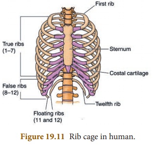
Sternum or Rib cage
Rib cage occupies the chest region.
It is a cone-shaped structure made up of Twelve pairs of ribs. Ribs are
attached to vertebrae at the back which curve around to form a cage. Ten pairs
of ribs are attached to the breast bone at the front. Two pairs of lower ribs
are free at front. These are called as free-floating ribs. Rib cage is set up
in such a way that it can contract and expand during the process of breathing.
Rib cage protects the underlying lungs, heart and some part of liver.
Humans and giraffes
have the same number of bones in the necks, but the vertebrae in a giraffe’s
neck are much, much larger.
II. Appendicular skeleton
The appendicular skeleton contains the bones in the appendages of the body, as well as the
structures that connect the appendages to the axial skeleton. Specifically, the
appendicular skeleton comprises the shoulder girdle; the arm, wrist, and hand
bones; the pelvic girdle; and the leg, ankle, and foot bones.
a.
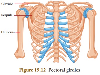
Shoulder bone or Pectoral bone
Shoulder bone is formed by collar
bone at the front and the shoulder blade at the back. The collar bone is
supported by breast bone at one end and the shoulder blade at the other end. The
shoulder bone encloses a socket like cavity into which fixes the ball of the
upper arm. This forms a ball and socket joint. This girdle is also called as
pectoral girdle.
b.
Pelvic bone
Pelvic bone is also called as pelvic
girdle. It is made up of strong bones to balance entire weight of the body.
Pelvic girdle is formed by five fused vertebrae at the back and form a cavity
in the centre while reaching the front part.
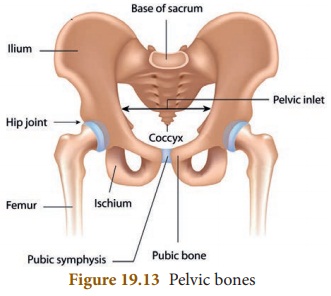
The thigh bones are attached to
either side of the girdle with a ball and socket joint.
c.
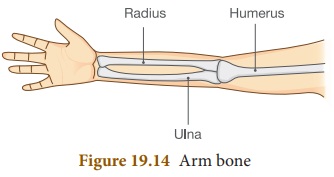
Arm bone
Arm bone is the upper limb made up
of humerus, radius, ulna, carpals, metacarpals and phalanges. All these bones
are joined by hinge joints which allow the limb to move only in one direction.
Humerus makes up the upper arm. Fore-arm is made up of radius and ulna. Wrist
is made up of carpals. Palm is made up of metacarpals. Fingers are made up of
phalanges.
d.
Leg bone
Leg bone is the lower limb made up
of femur, tibia, fibula, tarsals, metatarsals and phalanges. All these bones
are joined by hinge joints which allow the limb to move only in one direction.
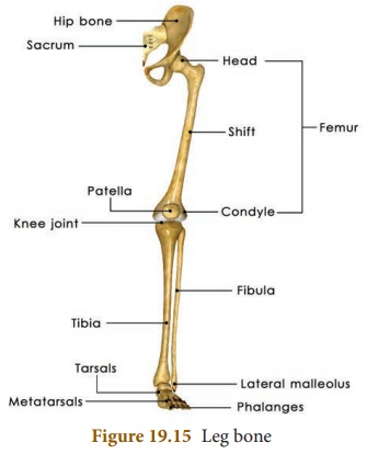
Knee is covered by a cap like
structure called as patella or a knee cap. Femur makes up the thigh bone. Leg
is made up of tibia and fibula. Ankle is made up of tarsals. Foot is made up of
metatarsals. Toes are made up of phalanges.
Related Topics