Chapter: Microbiology and Immunology: Bacteriology: Rickettsia, Orientia Ehrlichia, and Coxiella
Rickettsia rickettsiae
Rickettsia rickettsiae
R. rickettsiae causes Rocky Mountain spotted fever, the most seri-ous type of spotted fever. Rocky Mountain spotted fever was the first among the spotted fever group to be described. This dis-ease was earlier called as Mediterranean disease and later bou-tonneuse fever by Connor who described this condition for the first time in 1910. Megaw in 1917 first described this disease in the foothills of Himalaya in India.
Properties of the Bacteria
Morphology, culture, biochemical reactions, and other properties of R. rickettsiae are similar to those of other rick-ettsial pathogens. They are small intracellular bacteria, which multiply in the cytoplasm of the infected cells. They stain poorly with Gram stain, but stain well with Giemsa or Gimenez stain.
Pathogenesis and Immunity
R. rickettsiae like other rickettsiae multiply within the endo-thelial cells of the small blood vessels and invade the blood streams. Subsequently, they cause vasculitis and vascular lesions, which are found in almost all organs but are com-monly found in the skin and in the adrenal glands, liver, heart, and CNS. The condition progresses to hypoalbunemia, hyponatremia, and hypovolemia due to loss of plasma into the tissues.
Clinical Syndrome
R. rickettsiae cause Rocky Mountain spotted fever.
◗ Rocky mountain spotted fever
Incubation period is 7 days. The condition is characterized by development of fever, severe headache, chills, and myalgia. A rash may develop after three or more days and typically appears initially on wrist, ankles, and palms and soles and then spreads to the trunk. The rash is maculopapular early in the disease but may later become petechial and hemorrhagic.
This is a serious disease associated with many complica-tions, such as respiratory failure, encephalitis, and renal fail-ure. The patient may die within 5 days of onset of symptoms. The overall mortality rate is nearly 4% despite effective anti-biotic therapy. The deficiency of enzyme glucose-6-phosphate dehydrogenase (G6PD) is usually associated with more severity of infection.
Epidemiology
◗ Geographical distribution
Rocky Mountain spotted fever is distributed in southern Canada, Central America, Mexico, and parts of South America. R. rickettsiae, causing the condition, is the most common rick-ettsial pathogen in the United States.
◗ Habitat
R. rickettsiae is an intracellular pathogen of endothelial cellsand blood vessels of humans. The bacteria are also found in ticks.
◗ Reservoir, source, and transmission of infection
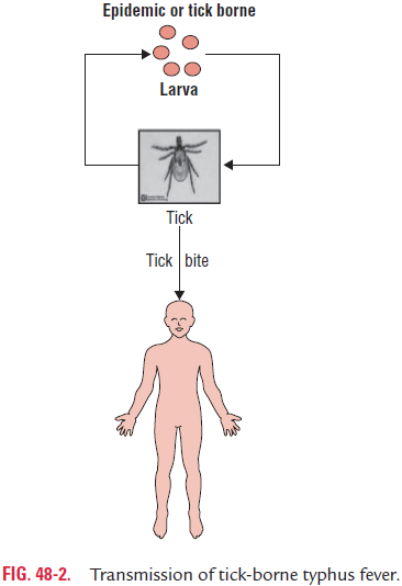
Ticks are the natural hosts, reservoirs, and vectors of R. rickettsiae. Different tick hosts are associated with theinfection in different parts of the world. These vectors include the wood tick (Dermacentor andersoni) in the western United States and western Canada, the American dog tick (Dermacentor variabilis) in the eastern Canada and eastern United States, and Lone star tick (Amblyomma americana) in the southwestern United States. Rickettsia multiplies in these ticks and is transmitted transovarially to the next gen-eration (Fig. 48-2).
R. rickettsiae is transmitted to humans through saliva duringthe bite by a tick. It usually takes 6 hours of attachment and feeding before rickettsiae are transmitted to the host. Infection can also be transmitted occasionally by scratching and rubbing infectious tick feces into the abraded skin.
Laboratory Diagnosis
◗ Specimens
These include skin biopsy for antigen detection and serum for serodiagnosis.
◗ Culture
R. rickettsiae can be isolated in embryonated egg or tissue cul-tures, but cultures are rarely attempted because of associated risk of infection.
◗ Direct detection of rickettsial antigen
Direct detection of R. rickettsiae in skin biopsy specimens of the rash from infected patients by direct fluorescent antibody test using the specific antirickettsial antibodies is a rapid and specific method for confirming diagnosis of Rocky Mountain spotted fever. This test is recommended for its use prior to therapy or within the first 48 hours after the antibiotic therapy.
◗ Serodiagnosis
Definite diagnosis of Rocky Mountain spotted fever is made by employing serological tests that detect R. rickettsiae immunoglobulin G (IgG) antibodies. IFA and enzyme linked immunosorbent assay (ELISA) are new serological tests that are used for early diagnosis of the condition.
The IFA is most commonly used test. This test uses group-specific heat-labile proteins and the LPS antigens of scrub typhus group. Therefore, the test is not species specific. The IFA is 95–100% sensitive and 100% specific. Demonstration of a fourfold rise in antibody titers between acute and convales-cent sera or demonstration of antibody titre of 1:64 or more in a single serum is diagnostic of the disease. These antibod-ies are detected in the serum 2–3 weeks after the onset of the disease and remain in the serum after a very long period of time.
Treatment
Tetracyclines, chloramphenicol, and fluoroquinolones, such as ciprofloxacin are effective against R. rickettsiae.
Prevention and Control
The use of protective clothing and insect repellents and avoid-ance of tick-infested areas are preventive measures against R.rickettsiae infection. No vaccine is available against the RockyMountain spotted fever.
Related Topics