Chapter: 11th 12th standard bio zoology Human Body higher secondary school
Reproduction of Riccia

Reproduction
The reproduction in Riccia takes place by means of (1) vegetative and (2) sexual methods.
Vegetative Reproduction
By the death and decay of the older parts of the thallus
By adventitious branches
By cell division or gemma formation
By tubers
By thick apices
By adventitious Branches
In several species of Riccia, the adventitious branches are produced on the ventral surface of the thallus. These branches get detached from the thalli and develop into new gametophytes.
By Tubers
In species such as R. discolor, the vegetative tubers develop at the apices of the branches of the thallus which face adverse conditions and develop into new plants on the approach of favourable conditions.
By cell division or gemma formation
Riccia may also reproduce vegetatively by cell division of the young rhizoids which develop into gemma-like structure. These structures give rise to new plants.
Sexual reproduction
Gemetophyte Generation
Majority of the species of Riccia are homothallic (monoecious) i.e., and antheridia and archegonia are borne upon the same thallus. The species of this type are R. glauca and R. gangetica. The heterothallic (dioecious) species are also common. In such species the antherdia and archegonia develop separately on different thalli and the important species of this type are R. himalayensisand R. frostii.
Sporophyte generation
At the time of formation of spores, there is genotypic type of sex determination i.e., two spores of a tetrad develop into female and two into male gametophytes, after meiosis.
Sexorgans
The sex organs, antheridia and archegonia, are produced in the groove situated on the dorsal surface of the mature gametophyte in acropetal succession.
In homothallic species, the alternate groups of antheridia and archegonia are found at a sufficient distance from the rowing point, even though the sex organs start growing initially on the floor of the dorsal groove as they grow, the surrounding issue also grows quickly. As a result of which a distinct chamber is seen surrounding the male and the female sex organs. They are the antheridial and archegonial chambers.
Structure of mature antheridium

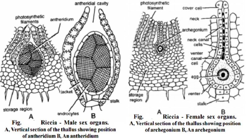
A mature antheridium remains embedded in an antheridial chamber, which opens by an ostiole on the dorsal side of the thallus. The mature antheridium consists of a few celled stalk and the antheridium proper. The antheridium proper may be rounded or somewhat pointed at its apical end. A sterile single layered jacket-layer encircles the antheridium and protects it. The mature antheridium contains androcytes within the jacket layer. Each androcyte metamorphoses into an antherozoid. Each antherozoid is a curved structure with two flagella.
Structure of mature archegonium
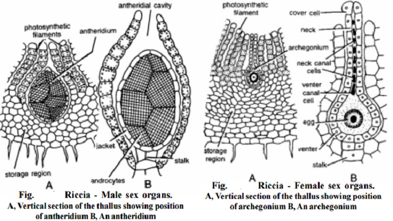
Mature archegonium is a flask-shaped structure attached to the thallus by a short stalk. It consists of an elongated neck made up of 6 rows of cells and a bulbous venter. The six vertical rows of the cells enclose a neck canal. There are four cover cells at the top of neck canal. Prior to maturity, the four neck canal cells, found within the neck canal, disintegrate into mucilaginous mass. The venter has a single layered wall around it which is of 12-20 cells in perimeter. The enter encloses a venter canal cell and the large egg. The venter canal cell disintegrates on maturity and only the large egg remains in the venter. The muilagenous mass absorbs water by imbibition and because of the pressure, the cover cells become separated from each other and an opening is formed. The mucilaginous mass extrudes through the opening and attracts the antherozoids, which enmass around the opening.
The venter cells are stimutated by the process of fertilization, they divide periclinally and the wall of the venter benomes two celled in thickness, ultimately a two layered calyptra is formed inside which, the developing embryo is situated.
Related Topics