Chapter: Pharmaceutical Drug Analysis: Theory and Technique of Quantitative Analysis
Radioimmunoassays (RIAS)
RADIOIMMUNOASSAYS (RIAS)
An assay method based on immunological antibody-hapten
(Ab-Ha) reaction that makes use of a radioactive tracer is usually known as
radioimmunoassay. A hapten (or haptene) is a small molecule that represents the
portion of an antigenic molecule or complex which determines its immunologic specificity,
for instance : cortisol ; whereas an antibody is a relatively large protein
that is specific for certain haptens. An antibody is generated by binding the
hapten to a protein, resulting into the formation of an antigen that
specifically stimulates the immune system to produce antibodies specific for
the hapten.
The assays that utilize protein instead of antibody are
normally termed as competitive protein
bind-ing assays. As an antibody is also a protein, therefore, a
radioimmunoassay may be looked upon as a type of competitive protein binding assay.
Theory :
Generally, a radioimmunoassay
makes use of a radioactive hapten and subsequently the percent of radioactivity bound to the antibody
is measured. The radioactivity is determined by the help of a Geiger-Müller
Counter or Geiger-Counter or G-M Tube and sometime by a Scintillation Counter.
First of all, a ‘Standard
Curve’ or a ‘Calibration Curve’
is plotted between the reciprocal value (i.e.,
1 × % –1 radioactivity bound to the antibody) versus the amount of
standard for a series of unknowns. Thus, the amount of hapten present in the
unknown sample is measured from the plotted calibration curve conveniently.
The radioimmunoassay is based
on the evolved competition between the combination of radioactive (Ha+)
and nonradioactive (Ha) hapten to the antibody as represented below :
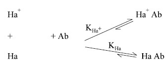
Let us assume that the binding
constants for Ha+ and Ha are equal ; now, for a fixed quantity of Ha+
but an increased concentration of Ha. The ultimate impact would be that lesser
Ha+ shall be bound. In actual practice, however, the use of Tritium
(H3) or Carbon-14 (C14), which helps to render the Ha
radioactive, ulti-mately maintains the equality of these binding constants,
namely : KHa+ and KHa . It also confirms that
the chemical properties of both radioactive (Ha+) and nonradioactive
(Ha) entities are more or less the same as far as the antibody is concerned.
Salient Features of Radioimmunoassays
·
They belong to a class of extremely sensitive methods of
analysis,
·
Sample required for assay is usually very small e.g., 1 ml of serum,
·
Concentrations upto the nanogram range i.e., 10–9 g can be measured
accurately,
·
A large number of hormones and drugs which find their
abundant usage in a bad way, namely :
·
cortisol (17-hydroxycorticosterone or hydrocortisone),
insulin, morphine, barbiturates (sedatives), vitamin B12, digoxin
and human growth hormones, such as : somatotropin (elaborated in the placenta),
·
Incidence of interferences observed in the radioimmunoassays
are fairly insignificant by virtue of the highly specific hapten-antibody
complexation reaction, and
·
Exceptions do occur when two 5-substituted barbiturates
present together cannot be assayed by this method, obviously due to
interference.
1. Cortisol (In Plasma)
Theory :
Cortisol (or hydrocortisone)
was introduced in the year 1951, for the treatment of rheumatoid arthritis. It has a significant effect
on protein metabolism. It also exerts widespread effects on carbohydrates,
lipid and protein synthesis (or anabolism). The cardinal side effects such as
excessive potassium excretion and sodium retention, enhanced gastric acidity,
oedema, psychosis and negative nitogen balance are some of the exaggerated
manifestations of the normal metabolite functions of cortisol.
Most importantly, the determination of cortisol levels is
considered useful in the diagnosis and treatment of various ailments, namely :
Addison’s Disease i.e., pernicious
anaemia—a condition whereby the maturation of the red cells may not proceed
beyond the stage of megaloblasts; Cushing’s Syndrome.
Adrenal Tumours :
The assay-method is entirely
based on the Schwartz-Mann Kit. According to this method, cortisol is first extracted from the plasma using CH2Cl2
(methylene chloride). In the actual radioimmunoassay the cortisol present in
the extract competes with Cortisol-H3 (i.e., the radioactive tracer) for the common binding sites on
transcortin, which is incidently not an antibody but a cortisol-binding
protein. Now, the free cortisol is quantitatively removed by adsorption on
dextran-coated charcoal from the one bound to the transcortin. Finally, the
bound radioactivity (due to Cortisol-H3) is measured which is then
employed to calculate exactly the amount of cortisol present in the sample by
the help of a Standard Curve (or Calibration Curve).
Materials Required :
Schwartz-Mann-H3 Cortisol RIA-Kit ; liquid
scintillation counter, centrifuge.
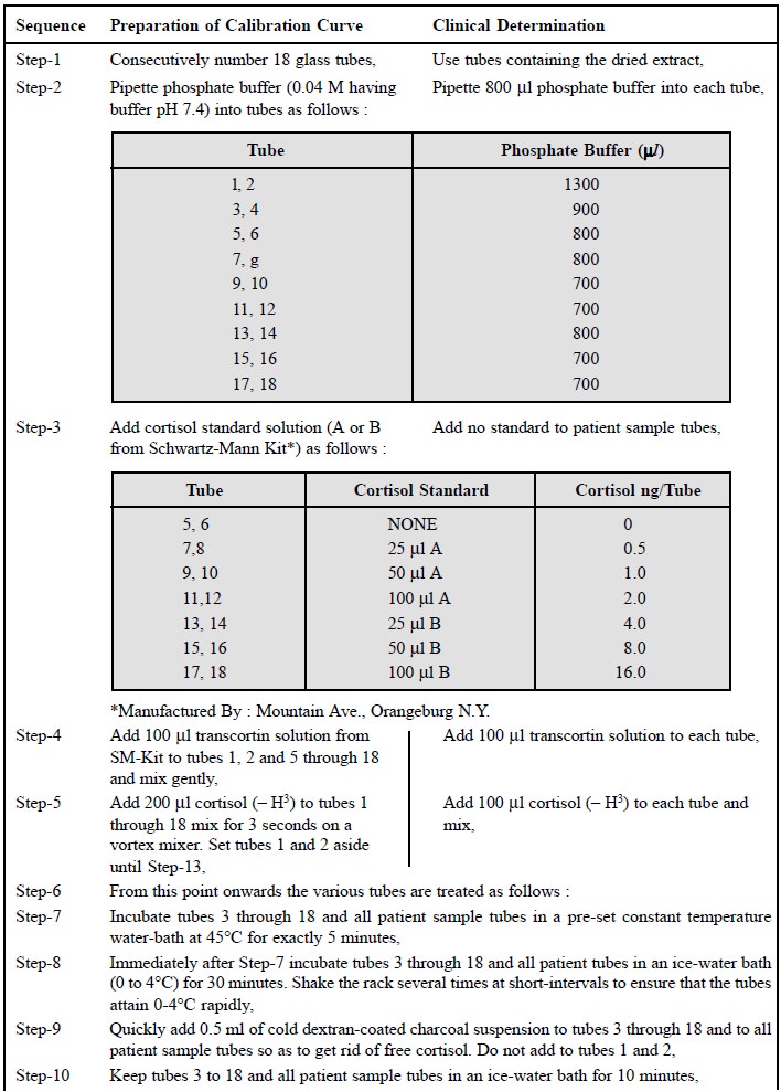
Procedure :
The various steps to be
followed sequentially for the assay of cortisol in plasma are as follows :
·
The cortisol is usually extracted from the samples drawn
from the patients, as follows :
Place 100 μ l of plasma in each of two
tubes and add 2.5 ml of methylene chloride. Shake the contents of the tube
vigorously for 10 minutes and transfer 0.5 ml of clear extract (i.e., the lower layer) to another tube.
Evaporate the methylene chloride either at 35°C in an oven or in a stream of N2.
The extract thus obtained is employed in the following step.
·
The following steps viz.,
Step 1 to Step 15, related to the procedure for the assay and the calibration
curves must be performed simultaneously :
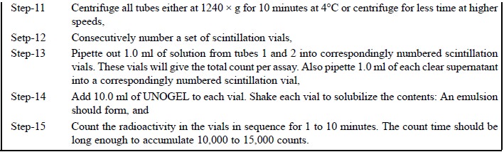
·
Results : Average the counts per minute
in vials 3 and 4. This is the blank value. Now, subtract the blank from all other counts per minute
to obtain the actual counts per minute and average the counts per minute for
vials 1 to 2 to find the total count per minute. The percent bound may be
calculated using the following expression :
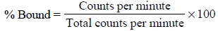
Finally, plot the percent bound
Vs nanograms (ng) per tube of
cortisol standard either on linear or on semilog paper and make use of this
Standard Curve to calculate the amount of cortisol present in the unkown
samples.
Related Topics