Chapter: 11th Botany : Chapter 6 : Cell: The Unit of Life
Plant Cell Organelles
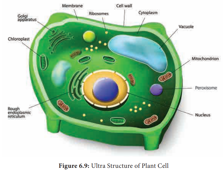
Cell Organelles
1. Endomembrane System
The system of membranes in a eukaryotic cell,
comprising the plasma membrane, nuclear membrane, endoplasmic reticulum, golgi
apparatus, lysosomes and vacuolar membranes (tonoplast). Endomembranes are made
up of phospholipids with embedded proteins that are similar to cell membrane
which occur within the cytoplasm. The endomembrane system is evolved from the
inward growth of cell membrane in the ancestors of the first eukaryotes (Figure
6.15).
2. Endoplasmic Reticulum
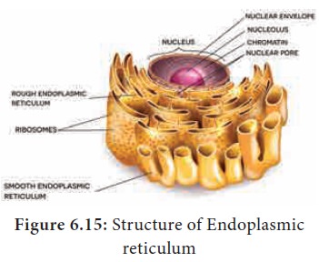
The largest of the internal membranes is called the endoplasmic reticulum (ER). The name endoplasmic reticulum was given by K.R. Porter (1948). It consists of double membrane. Morphologically the structure of endoplasmic reticulum consists of:
1
Cisternae
are long,
broad, flat, sac like structures
arranged in parallel bundles or stacks to form lamella. The space between
membranes of cisternae is filled with fluid.
2
Vesicles are oval
membrane bound vacuolar structure.
3
Tubules are
irregular shape, branched, smooth
walled, enclosing a space
Endoplasmic reticulum is associated with nuclear
membrane and cell surface membrane. It forms a network in cytoplasm and gives
mechanical support to the cell. Its chemical environment enables protein
folding and undergo modification necessary for their function. Misfolded
proteins are pulled out and are degraded in endoplasmic reticulum. When
ribosomes are present in the outer surface of the membrane it is called as rough endoplasmic reticulum(RER), when the ribosomes are absent in the
endoplasmic reticulum it is called as smooth
Endoplasmic reticulum(SER).
Rough endoplasmic reticulum is
involved in protein synthesis and smooth endoplasmic reticulum are the sites of
lipid synthesis. The smooth endoplasmic reticulum contains enzymes that
detoxify lipid soluble drugs, certain chemicals and other harmful compounds.
3. Golgi Body (Dictyosomes)
In 1898, Camillo
Golgi visualized a netlike reticulum of fibrils near the nucleus, were
named as Golgi bodies. In plant
cells they are found as smaller vesicles termed as dictyosomes. Golgi apparatus is a stack of flat membrane enclosed sacs. It consist of cisternae, tubules,
vesicles and golgi vacuoles. In plants the cisternae are 10-20 in number placed
in piles separated from each other by a thin layer of inter cisternal cytoplasm
often flat or curved. Peripheral edge of cisternae forms a network of tubules
and vesicles. Tubules interconnect cisternae and are 30-50nm in dimension.
Vesicles are large round or concave sac. They are pinched off from the
tubules.They are smooth/secretary or coated type. Golgi vacuoles are large
spherical filled with granular or amorphous substance, some function like
lysosomes. The Golgi apparatus compartmentalises a series of steps leading to
the production of functional protein.
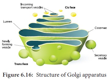
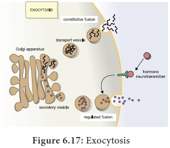
Small pieces of rough endoplasmic reticulum are
pinched off at the ends to form small vesicles. A number of these vesicles then
join up and fuse together to make a Golgi body. Golgi complex plays a major
role in post translational modification of proteins and glycosidation of lipids
(Figure 6.16 and 6.17).
Functions:
·
Glycoproteins and glycolipids are produced
·
Transporting and storing lipids.
· Formation of lysosomes.
·
Production of digestive enzymes.
·
Cell plate and cell wall formation
·
Secretion of Carbohydrates for the formation of
plant cell walls and insect cuticles.
· Zymogen granules (proenzyme/pre-cursor of all enzyme) are synthesised.
4. Mitochondria
It was first observed by A. Kolliker (1880). Altmann (1894)
named it as Bioplasts. Later Benda (1897,
1898), named as mitochondria. They
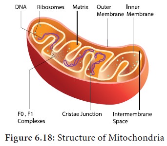
are ovoid, rounded, rod shape and pleomorphic structures. Mitochondrion
consists of double membrane, the outer and inner membrane. The outer membrane
is smooth, highly permeable to small molecules and it contains proteins called Porins, which form channels that allows
free diffusion of molecules smaller than about 1000 daltons and the inner
membrane divides the mitochondrion into two compartments, outer chamber between
two membranes and the inner chamber filled with matrix.
The inner membrane is convoluted (infoldings),
called crista (plural: cristae).
Cristae contain most of the enzymes for electron transport system. Inner
chamber of the mitochondrion is filled with protein aceous material called mitochondrial matrix. The inner
membrane consists of stalked particles called elementary particles or Fernandez
Moran particles, F1 particles or
Oxysomes. Each particle consists of
a base, stem and a round head. In the head ATP synthase is present for
oxidative phosphorylation. Inner membrane is impermeable to most ions, small
molecules and maintains the proton gradient that drives oxidative
phosphorylation (Figure 6.18).
Mitochondria contain 73% of proteins, 25- 30% of
lipids, 5-7 % of RNA, DNA (in traces) and enzymes (about 60 types).
Mitochondria are called Power house of a
cell, as they produce energy rich
ATP.
All the enzymes of Kreb’s cycle are found in the
matrix except succinate dehydrogenase. Mitochondria consist of circular DNA and
70S ribosome. They multiply by fission and replicates by strand displacement
model. Because of the presence of DNA it is semi-autonomous organelle. Unique
characteristic of mitochondria is that they are inherited from female parent
only. Mitochondrial DNA comparisons are used to trace human origins.
Mitochondrial DNA is used to track and date recent evolutionary time because it
mutates 5 to 10 time faster than DNA in the nucleus.
5. Plastids
The term plastid is derived from the Greek word Platikas
(formed/moulded) and used by A.F.U.
Schimper in 1885. He classified plastids into following types according to
their structure, pigments and function. Plastids multiply by fission.
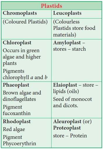
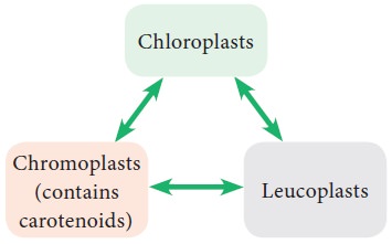
According to Schimper, different kind of plastids
can transform into one another.
6. Chloroplast
Chloroplasts are vital organelle found in green plants. Chloroplast has a double membrane the outer membrane and the inner membrane separated by a space called periplastidial space. The space enclosed by the inner membrane of chloroplast is filled with gelatinous matrix, lipo-proteinaceous fluid called stroma. Inside the stroma there is flat interconnected sacs called thylakoid. The membrane of thylakoid enclose a space called thylakoid lumen.
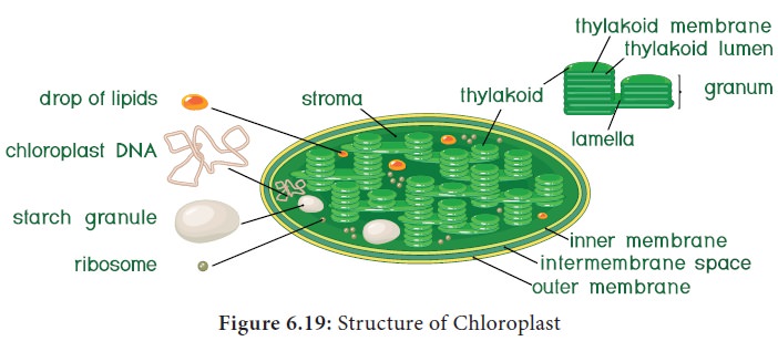
Grana (singular:
Granum) are formed when many of
these thylakoids are stacked together like pile of coins. Light is absorbed and
converted into chemical energy in the granum, which is used in stroma to
prepare carbohydrates. Thylakoid contain chlorophyll pigments. The chloroplast
contains osmophilic granules, 70s ribosomes, DNA (circular and non histone) and
RNA. These chloroplast genome encodes approximately 30 proteins involved in
photosynthesis including the components of photosystem I & II, cytochrome
bf complex and ATP synthase. One of the subunits of Rubisco is encoded by
chloroplast DNA. It is the major protein component of chloroplast stroma,
single most abundant protein on earth. The thylakoid contain small, rounded
photosynthetic units called quantosomes.
It is a semi-autonomous organelle and divides by fission (Figure 6.19).
Functions:
•
Photosynthesis
•
Light reactions takes place in granum,
•
Dark reactions take place in stroma,
•
Chloroplast is involved in photo respiration.
7. Ribosome
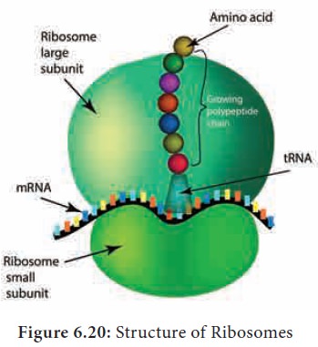
Ribosomes were first observed by George Palade (1953) as dense particles or granules in the electron microscope. Electron microscopic observation
reveals that ribosomes are composed of two rounded sub units, united together
to form a complete unit. Mg2+ is required for structural cohesion of ribosomes.
Biogenesis of ribosome are denova formation,
auto replication and nucleolar
origin. Each ribosome is made up of one small and one large sub-unit Ribosomes
are the sites of protein synthesis in the cell. Ribosome is not a membrane
bound organelle (Figure 6.20).
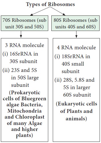
Ribosome consists of RNA and
protein: RNA 60 % and Protein 40%. During protein synthesis many ribosomes are
attached to the single mRNA is called polysomes
or polyribosomes. The function of polysomes is the formation
of several copies of a particular polypeptide during protein synthesis. They
are free in non-protein synthesising cells. In protein synthesising cells they
are linked together with the help of Mg2+ ions.
8. Lysosomes (Suicidal Bags of Cell)
Lysosomes were discovered by Christian de Duve (1953),
these are known as suicidal bags.
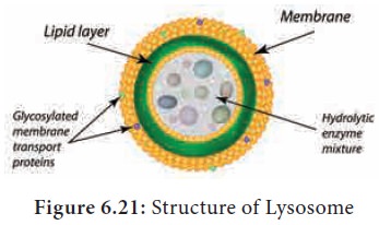
They are spherical bodies enclosed
by a single unit membrane. They are found in eukaryotic cell. Lysosomes are
small vacuoles formed when small pieces of golgi body are pinched off from its
tubules.
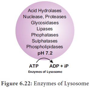
They contain a variety of hydrolytic enzymes, that
can digest material within the cell. The membrane around lysosome prevent these
enzymes from digesting the cell itself (Figure 6.21).
Functions:
·
Intracellular
digestion: They digest carbohydrates,
proteins and lipids present in cytoplasm.
·
Autophagy:
During
adverse condition they digest their
own cell organelles like mitochondria and endoplasmic reticulum
![]()
![]()
![]()
·
Autolysis:
Lysosome
causes self destruction of cell on
insight of disease they destroy the cells.
·
Ageing: Lysosomes
have autolytic enzymes that disrupts
intracellular molecules.
·
Phagocytosis:
Large
cells or contents are engulfed and
digested by macrophages, thus forming a phagosome in cytoplasm. These phagosome
fuse with lysosome for further digestion.
·
Exocytosis:
Lysosomes
release their enzymes outside the
cell to digest other cells (Figure 6.22).
9. Peroxisomes
Peroxisomes were identified as
organelles by Christian de Duve
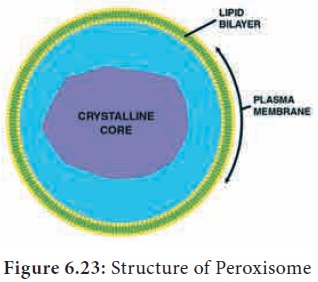
(1967). Peroxisomes are small spherical bodies and single membrane bound
organelle. It takes part in photorespiration and associated with glycolate
metabolism. In plants, leaf cells have many peroxisomes. It is also commonly
found in liver and kidney of mammals. These are also found in cells of protozoa
and yeast (Figure 6.23).
10. Glyoxysomes
Glyoxysome was discovered by Harry Beevers (1961).
Glyoxysome is a single membrane
bound organelle. It is a sub cellular organelle and contains enzymes of
glyoxylate pathway. β-oxidation of fatty acid occurs in glyoxysomes of
germinat-ing seeds Example: Castor seeds.
11. Microbodies
Eukaryotic cells contain many enzyme bearing
membrane enclosed vesicles called microbodies.
They are single unit membrane bound cell organelles: Example: peroxisomes and
glyoxysomes.
12. Sphaerosomes
It is spherical in shape and enclosed by single
unit membrane. Example: Storage of fat in the endosperm cells of oil seeds.
13. Centrioles
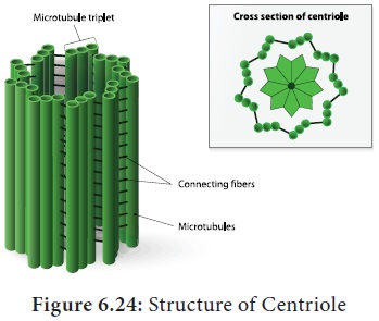
Centriole consist of nine triplet peripheral
fibrils made up of tubulin. The central part of the centriole is called hub, is connected to the tubules of the
peripheral triplets by radial spokes (9+0 pattern). The centriole form the
basal body of cilia or flagella and spindle fibers which forms the spindle
apparatus in animal cells. The membrane is absent in centriole (non-membranous
organelle) (Figure 6.24).
14. Vacuoles
In plant cells vacuoles are large, bounded by a
single unit membrane called Tonoplast.
The vacuoles contain cell sap, which is a solution of sugars, amino acids,
mineral salts, waste chemical and anthocyanin pigments. Beetroot cells contains
anthocyanin pigments in their vacuoles. Vacuoles accumulate products like
tannins. The osmotic expansion of a cell kept in water is chiefly regulated by
vacuole and the water enters the vacuoles by osmosis.
The major function of plant vacuole is to maintain
water pressure known as turgor pressure
, which maintains the plant structure. Vacuoles organises itself into a
storage/sequestration compartment. Example: Vacuoles store, most of the sucrose
of the cell.
i. Sugar in Sugar
beet and Sugar cane.
ii. Malic acid in Apple.
iii. Acids in Citrus
fruits.
iv. Flavonoid pigment Cyanidin 3 rutinoside in the petals of Antirrhinum.
v. Tannins in Mimosa pudica.
vi. Raphide crystals in Dieffenbachia.
vii. Heavy metals in Mustard (Brassica).
viii. Latex in Rubber tree and Dandelion stem.
Cell Inclusions
The cell inclusions are the non-living materials
present in the cytoplasm. They are organic and inorganic compounds.
Inclusions in prokaryotes
In prokaryotes, reserve materials such as phosphate
granules, cyanophycean granules, glycogen granules, poly β-hydroxy butyrate
granules, sulphur granules, carboxysomes and gas vacuoles are present.
Inorganic inclusions in bacteria are polyphosphate granules (volutin granules)
and sulphur granules. These granules are also known as metachromatic granules.
Inclusions in Eukaryotes
•
Reserve food materials: Starch grains, glycogen
granules, aleurone grains, fat droplets
•
Secretions in plant cells are essential oil, resins,
gums, latex and tannins
•
Inorganic
crystals – plant cell have calcium
carbonate, calcium oxalate and silica
•
Cystolith
–
hypodermal leaf cells of Ficus bengalensis, calcium carbonate
•
Sphaeraphides
– star
shaped calcium oxalate, Colocasia
•
Raphides – calcium
oxalate, Eichhornia
•
Prismatic
crystals – calcium oxalate, dry scales
of Allium cepa
Related Topics