Chapter: 11th Botany : Chapter 6 : Cell: The Unit of Life
Electron Microscope
Electron Microscope
Electron Microscope was first introduced by Ernest Ruska (1931) and developed by G Binning and H Roher (1981). It is used to analyse the fine details of the cell
and organelles called ultrastructure. It uses beam of accelerated electrons as
source of illumination and therefore the resolving power is 1,00,000 times than
that of light microscope.
The specimen to be viewed under electron microscope
is dehydrated and impregnated with electron opaque chemicals like gold or
palladium. This is essential for withstanding electrons and also for contrast of the image.
There are two kinds of electron microscopes namely
1.
Transmission Electron Microscope (TEM)
2.
Scanning Electron Microscope (SEM)
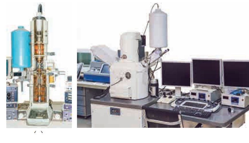
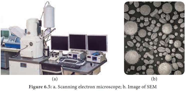
1. Transmission electron microscope
Transmission
electron microscope: This is the most commonly used electron microscope
which provides two dimensional image. The components of the microscope are as
follows:
![]()
![]()
![]()
a.
Electron Generating System
b.
Electron Condensor
c.
Specimen Objective
d.
Tube Lens
e.
Projector
A beam of electron passes through the specimen to
form an image on fluorescent screen. The magnification is 1–3 lakhs times and
resolving power is 2–10 Å. It is used for studying detailed structrue of
viruses, mycoplasma, cellular organelles, etc (Figure 6.4 a and b).
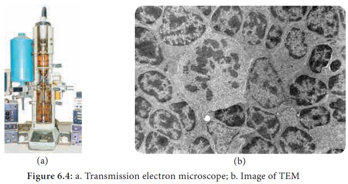
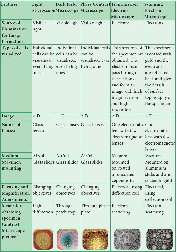
2. Scanning Electron Microscope:
This is used to obtain three dimensional image and
has a lower resolving power than TEM. In this, electrons are focused by means
of lenses into a very fine point. The interaction of electrons with the
specimen results in the release of different forms of radiation (such as auger
electrons, secondary electrons, back scattered electrons) from the surface of
the specimen. These radiations are then captured by an appropriate detector,
amplified and then imaged on fluorescent screen. The magnification is 2,00,000
times and resolution is 5–20 nm (Figure 6.5 a and b).
Comparison of Microscopes
Related Topics