Chapter: Anatomy of Flowering Plants: An Introduction to Structure and Development : Flower
Ovule in Flower
Ovule
Ovule
primordia are initiated as small swellings in the placenta. The ovule consists
of a nucellus, which bears the embryo sac, enclosed by one or two (inner and
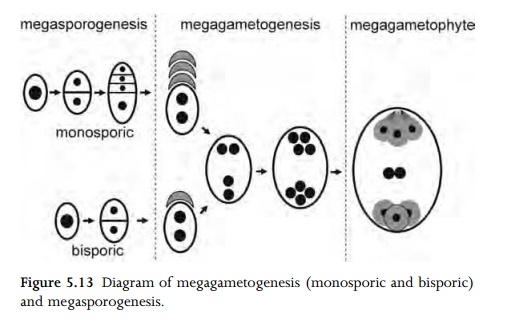
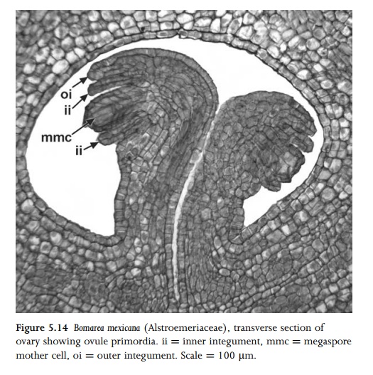
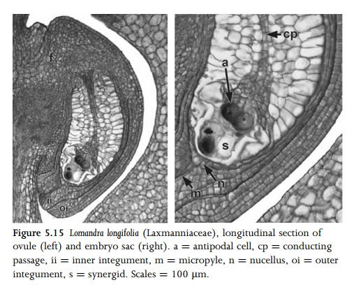
outer) integuments (Figs 5.13 5.15). The region where the nucellus, integuments
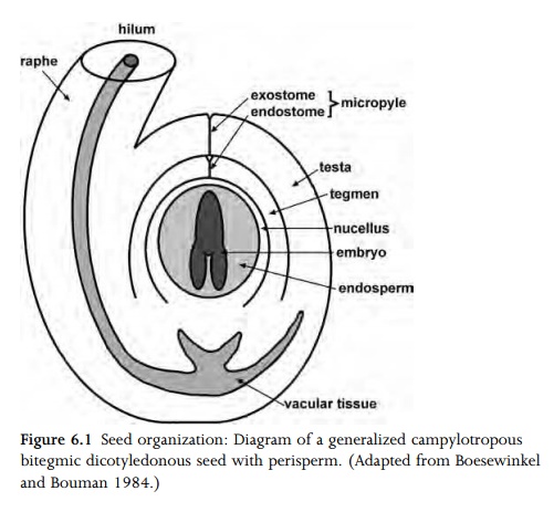
and funicle unite is termed the chalaza (Fig. 6.1). The micropyle is a narrow
opening in the ovule formed by one or both integuments, located at the opposite
end of the embryo sac to the chalaza. At anthesis, each ovule is attached to
the ovary by a funicle, which normally possesses a single vascular strand. The
vascular bundle in the funicle usually terminates at the chalaza, but in some
species it is more extensive.
The
nucellus arises from the apex and body of the ovule primordium. The integuments
develop from around the primor-dium base and encircle its apex, forming the
micropyle; normally the inner integument is initiated before the outer. The
possession of two integuments is the most common condition in angio-sperms, but
a single integument characterizes some eudicots, and a few species lack
integuments entirely16. Prior to formation of the archespore, the
ovule primordium is organized into either two or (more commonly) three distinct
zones defined by orienta-tion of cell division. The outermost (dermal) layer
initially undergoes mainly anticlinal divisions, but may later proliferate at
the micropylar end to form a nucellar cap, and sometimes also proliferates
around and below the embryo sac. The archespore forms in the subdermal layer, immediately
subtended by the central zone.
In some
species the nucellus proliferates in the central zone to form a hypostase,
which is a chalazal region of often refractive and suberinized or lignified
tissue. For example, a large hypostase is present in Acorus and Crocus.
Sometimes other specialized structures are formed in the central zone of the
nucellus, such as a postament or conducting passage (Fig. 5.15); these often
persist after fertilization. The dermal nucellar cells surrounding the embryo
sac often break down before (or shortly after) fertili-zation, in which case
the innermost epidermal layer of the inner integument is sometimes
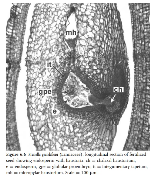
differentiated to form an endo-thelium or integumentary tapetum (Fig. 6.6).
Endothelium cells are frequently enlarged and densely cytoplasmic, and
sometimes endopolyploid; in some species they have a secretory function.
Related Topics