Chapter: Ophthalmology: Ocular Trauma
Ocular Trauma: Examination Methods
Ocular Trauma
Examination Methods
The incidence of ocular injuries remains high
despite the increase in safety regulations in recent years, such as mandatory
seat belts and protective eye-wear for persons operating high-speed rotary
machinery. Therefore it is important that every general practitioner and health
care staff member is able to recognize an ocular injury and provide initial
treatment. The patient should then be referred to an ophthalmologist, who
should be solely responsible for evaluation of the injury and definitive
treatment. The follow-ing diagnostic options are available to determine the
nature of the injury more precisely.
Patient history:
Obtaining a thorough history will provide important infor-mation
about the cause of the injury.
âť– Work with a hammer and chisel nearly always
suggests an intraocular for-eign body.
âť– Cutting and grinding work suggests corneal
foreign bodies.
âť–
Welding and flame cutting work suggests ultraviolet
keratoconjunctivitis.
The examiner should always ascertain whether
the patient has ade-quate tetanus immunization.
Inspection (gross morphologic examination):
Ocular injuries frequentlycause
pain, photophobia, and blepharospasm. A few drops of topical anes-thetic are
recommended to allow the injured eye to be examined at rest with minimal pain
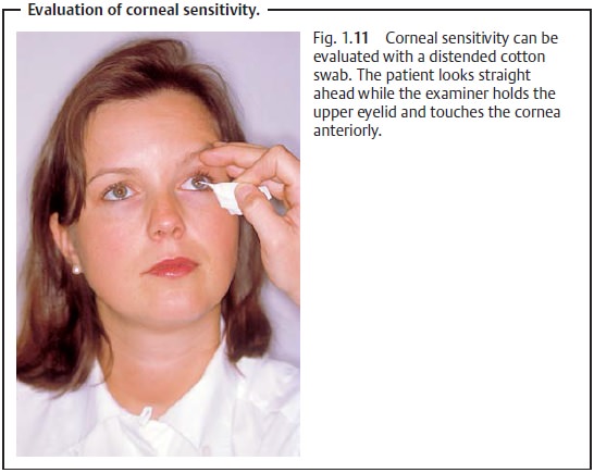
to the patient. The cornea and conjunctiva are then examined for signs of
trauma using a focused light, preferably one combined with a magnifying loupe
(see Fig. 1.11 for examination
technique). The eyelids may be everted to inspect the tarsal surface and
conjunctival fornix. A foreign body can then be removed immediately.
Ophthalmoscopy:
Examination with a focused light or ophthalmoscope willpermit
gross evaluation of deeper intraocular structures, such as whether a vitreous
or retinal hemorrhage is present. A vitreous hemorrhage may be identified by
the lack of red reflex on retroillumination. Care should be taken to avoid
unnecessary manipulation of the eye in an obviously severe open-globe injury (characterized by a soft globe,
pupil displaced toward the pene-tration site, prolapsed iris, and intraocular
bleeding in the anterior chamber and vitreous body). Such manipulation might
otherwise cause further dam-age, such as extrusion of intraocular contents.
To properly estimate the urgency of treating
palpebral and ocular trauma, it is particularly important to differentiate
between open-globe injuries and closed-globe injuries. Open-globe injuries have
highest priority due to the risk of losing the eye.
Related Topics