Chapter: Medical Physiology: Muscle Blood Flow and Cardiac Output During Exercise; the Coronary Circulation and schemic Heart Disease
Normal Coronary Blood Flow

Normal Coronary Blood Flow
The resting coronary blood flow in the human being averages about 225 ml/min, which is about 4 to 5 per cent of the total cardiac output.
During strenuous exercise, the heart in the young adult increases its cardiac output fourfold to seven-fold, and it pumps this blood against a higher than normal arterial pressure. Consequently, the work

output of the heart under severe conditions may increase sixfold to ninefold. At the same time, the coronary blood flow increases threefold to fourfold to supply the extra nutrients needed by the heart. This increase is not as much as the increase in workload which means that the ratio of energy expenditure by the heart to coronary blood flow increases. Thus, the efficiency” of cardiac utilization of energy increases to make up for the relative deficiency of coronary blood supply.
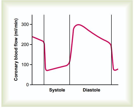
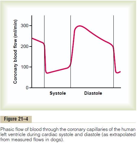
Phasic Changes in Coronary Blood Flow During Systole and Diastole—Effect of Cardiac Muscle Compression. Figure21–4 shows the changes in blood flow through thenutrient capillaries of the left ventricular coronarysystem in milliliters per minute in the human heart during systole and diastole, as extrapolated from experiments in lower animals. Note from this diagram that the coronary capillary blood flow in the left ven-tricle muscle falls to a low value during systole, which is opposite to flow in vascular beds elsewhere in the body. The reason for this is strong compression of the left ventricular muscle around the intramuscular vessels during systolic contraction.

During diastole, the cardiac muscle relaxes and no longer obstructs blood flow through the left ventricu-lar muscle capillaries, so that blood flows rapidly during all of diastole.
Blood flow through the coronary capillaries of the right ventricle also undergoes phasic changes duringthe cardiac cycle, but because the force of contraction of the right ventricular muscle is far less than that of the left ventricular muscle, the inverse phasic changes are only partial in contrast to those in the left ventric-ular muscle.

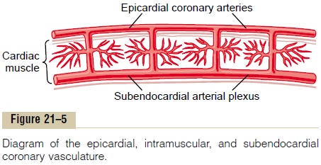
Epicardial Versus Subendocardial Coronary Blood Flow—Effect of Intramyocardial Pressure. Figure 21–5 demonstrates the special arrangement of the coronary vessels at dif- ferent depths in the heart muscle, showing on the outer surface epicardial coronary arteries that supply most of the muscle. Smaller, intramuscular arteries derived from the epicardial arteries penetrate the muscle, supplying the needed nutrients. Lying immediately beneath the endocardium is a plexus of subendocar- dial arteries. During systole, blood flow through the subendocardial plexus of the left ventricle, where the intramuscular coronary vessels are compressed greatly by ventricular muscle contraction, tends to be reduced. But the extra vessels of the subendocardial plexus nor- mally compensate for this. Later, we will see that this peculiar difference between blood flow in the epicardial and subendocardial arteries plays an important role in certain types of coronary ischemia.
Related Topics