Chapter: Medical Physiology: Muscle Blood Flow and Cardiac Output During Exercise; the Coronary Circulation and schemic Heart Disease
Coronary Circulation
Coronary Circulation
About one third of all deaths in the affluent society of the Western world result from coronary artery disease, and almost all elderly people have at least some impairment of the coronary artery circulation. For this reason, understanding normal and pathological physi-ology of the coronary circulation is one of the most important subjects in medicine.
Physiologic Anatomy of the Coronary Blood Supply
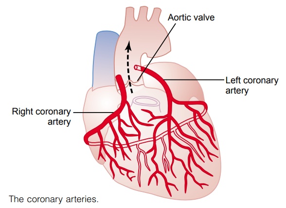
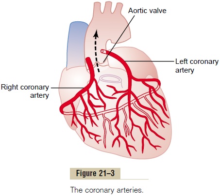
Figure 21–3 shows the heart and its coronary blood supply. Note that the main coronary arteries lie on the surface of the heart and smaller arteries then pene-trate from the surface into the cardiac muscle mass. It is almost entirely through these arteries that the heart receives its nutritive blood supply. Only the inner 1/10 millimeter of the endocardial surface can obtain sig-nificant nutrition directly from the blood inside the cardiac chambers, so that this source of muscle nutri-tion is minuscule.
The left coronary artery supplies mainly the anterior and left lateral portions of the left ventricle, whereas the right coronary artery supplies most of the right ventricle as well as the posterior part of the left ven-tricle in 80 to 90 per cent of people.
Most of the coronary venous blood flow from the left ventricular muscle returns to the right atrium of the heart by way of the coronary sinus—which is about 75 per cent of the total coronary blood flow. And most of the coronary venous blood from the right ventricu-lar muscle returns through small anterior cardiac veins that flow directly into the right atrium, not by way of the coronary sinus. A very small amount of coronary venous blood also flows back into the heart through very minute thebesian veins, which empty directly into all chambers of the heart.
Normal Coronary Blood Flow
The resting coronary blood flow in the human being averages about 225 ml/min, which is about 4 to 5 per cent of the total cardiac output.
During strenuous exercise, the heart in the young adult increases its cardiac output fourfold to seven-fold, and it pumps this blood against a higher than normal arterial pressure. Consequently, the work
output of the heart under severe conditions may increase sixfold to ninefold. At the same time, the coronary blood flow increases threefold to fourfold to supply the extra nutrients needed by the heart. This increase is not as much as the increase in workload which means that the ratio of energy expenditure by the heart to coronary blood flow increases. Thus, the efficiency” of cardiac utilization of energy increases to make up for the relative deficiency of coronary blood supply.
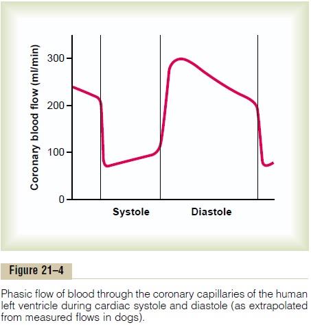
Phasic Changes in Coronary Blood Flow During Systole and Diastole—Effect of Cardiac Muscle Compression. Figure 21–4 shows the changes in blood flow through thenutrient capillaries of the left ventricular coronarysystem in milliliters per minute in the human heart during systole and diastole, as extrapolated from experiments in lower animals. Note from this diagram that the coronary capillary blood flow in the left ven-tricle muscle falls to a low value during systole, which is opposite to flow in vascular beds elsewhere in the body. The reason for this is strong compression of the left ventricular muscle around the intramuscular vessels during systolic contraction.
During diastole, the cardiac muscle relaxes and no longer obstructs blood flow through the left ventricu-lar muscle capillaries, so that blood flows rapidly during all of diastole.
Blood flow through the coronary capillaries of the right ventricle also undergoes phasic changes duringthe cardiac cycle, but because the force of contraction of the right ventricular muscle is far less than that of the left ventricular muscle, the inverse phasic changes are only partial in contrast to those in the left ventricular muscle.
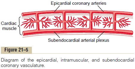
Epicardial Versus Subendocardial Coronary Blood Flow—Effect of Intramyocardial Pressure. Figure 21–5 demonstrates the special arrangement of the coronary vessels at dif- ferent depths in the heart muscle, showing on the outer surface epicardial coronary arteries that supply most of the muscle. Smaller, intramuscular arteries derived from the epicardial arteries penetrate the muscle, supplying the needed nutrients. Lying immediately beneath the endocardium is a plexus of subendocar- dial arteries. During systole, blood flow through the subendocardial plexus of the left ventricle, where the intramuscular coronary vessels are compressed greatly by ventricular muscle contraction, tends to be reduced. But the extra vessels of the subendocardial plexus nor- mally compensate for this. Later, we will see that this peculiar difference between blood flow in the epicardial and subendocardial arteries plays an important role in certain types of coronary ischemia.
Control of Coronary Blood Flow
Local Muscle Metabolism Is the Primary Controller of Coronary Flow
Blood flow through the coronary system is regulated mostly by local arteriolar vasodilation in response to cardiac muscle need for nutrition. That is, whenever the vigor of cardiac contraction is increased, regard-less of cause, the rate of coronary blood flow also increases. Conversely, decreased heart activity is accompanied by decreased coronary flow. This local regulation of coronary blood flow is almost identical to that occurring in many other tissues of the body, especially in the skeletal muscles all over the body.
Oxygen Demand as a Major Factor in Local Coronary Blood Flow Regulation. Blood flow in the coronaries usually is reg-ulated almost exactly in proportion to the need of the cardiac musculature for oxygen. Normally, about 70 per cent of the oxygen in the coronary arterial blood is removed as the blood flows through the heart muscle. Because not much oxygen is left, very little additional oxygen can be supplied to the heart mus-culature unless the coronary blood flow increases. For-tunately, the coronary blood flow does increase almost in direct proportion to any additional metabolic con-sumption of oxygen by the heart.
However, the exact means by which increased oxygen consumption causes coronary dilation has not been determined. It is speculated by many research workers that a decrease in the oxygen concentration in the heart causes vasodilator sub-stances to be released from the muscle cells and that these dilate the arterioles. A substance with great vasodilator propensity is adenosine. In the presence of very low concentrations of oxygen in the muscle cells, a large proportion of the cell’s ATP degrades to adeno-sine monophosphate; then small portions of this are further degraded and release adenosine into the tissue fluids of the heart muscle, with resultant increase in local coronary blood flow. After the adenosine causes vasodilation, much of it is reabsorbed into the cardiac cells to be reused.
Adenosine is not the only vasodilator product that has been identified. Others include adenosine phosphate compounds, potassium ions, hydrogen ions, carbon dioxide, bradykinin, and, possibly, prostaglandins and nitric oxide.
Yet, difficulties with the vasodilator hypothesis exist. First, pharmacologic agents that block or partially block the vasodilator effect of adenosine do not prevent coronary vasodilation caused by increased heart muscle activity. Second, studies in skeletal muscle have shown that continued infusion of adeno-sine maintains vascular dilation for only 1 to 3 hours, and yet muscle activity still dilates the local blood vessels even when the adenosine can no longer dilate them. Therefore, the other vasodilator mechanisms listed above must be remembered.
Nervous Control of Coronary Blood Flow
Stimulation of the autonomic nerves to the heart can affect coronary blood flow both directly and indirectly. The direct effects result from action of the nervous transmitter substances acetylcholine from the vagus nerves and norepinephrine and epinephrine from the sympathetic nerves on the coronary vessels them-selves. The indirect effects result from secondary changes in coronary blood flow caused by increased or decreased activity of the heart.
The indirect effects, which are mostly opposite to the direct effects, play a far more important role in normal control of coronary blood flow. Thus, sympa-thetic stimulation, which releases norepinephrine and epinephrine, increases both heart rate and heart con-tractility as well as increases the rate of metabolism of the heart. In turn, the increased metabolism of the heart sets off local blood flow regulatory mechanisms for dilating the coronary vessels, and the blood flow increases approximately in proportion to the meta-bolic needs of the heart muscle. In contrast, vagal stim-ulation, with its release of acetylcholine, slows the heart and has a slight depressive effect on heart con-tractility. These effects in turn decrease cardiac oxygen consumption and, therefore, indirectly constrict the coronary arteries.
Direct Effects of Nervous Stimuli on the Coronary Vasculature.
The distribution of parasympathetic (vagal) nerve fibers to the ventricular coronary system is not very great. However, the acetylcholine released by parasym-pathetic stimulation has a direct effect to dilate the coro-nary arteries.
There is much more extensive sympathetic innerva-tion of the coronary vessels. We see that the sympathetic transmitter substances norepineph-rine and epinephrine can have either vascular con-strictor or vascular dilator effects, depending on the presence or absence of constrictor or dilator receptors in the blood vessel walls. The constrictor receptors are called alpha receptors and the dilator receptors are calledbeta receptors. Both alpha and beta receptors exist in the coronary vessels. In general, the epicardial coronary vessels have a preponderance of alpha recep-tors, whereas the intramuscular arteries may have a preponderance of beta receptors. Therefore, sympa-thetic stimulation can, at least theoretically, cause slight overall coronary constriction or dilation, but usually constriction. In some people, the alpha vaso-constrictor effects seem to be disproportionately severe, and these people can have vasospastic myocar-dial ischemia during periods of excess sympathetic drive, often with resultant anginal pain.
Metabolic factors—especially myocardial oxygen consumption—are the major controllers of myocardial blood flow. Whenever the direct effects of nervous stimulation alter the coronary blood flow in the wrong direction, the metabolic control of coronary flow usually overrides the direct coronary nervous effects within seconds.
Related Topics