Geographical Distribution, Habitat, Morphology, Life Cycle, Pathogenesis, Clinical Manifestations, Laboratory Diagnosis, Treatment, Prevention and Control - Nematode: Ascaris Lumbricoides | 12th Microbiology : Chapter 8 : Medical Parasitology
Chapter: 12th Microbiology : Chapter 8 : Medical Parasitology
Nematode: Ascaris Lumbricoides
Nematode: Ascaris
Lumbricoides
Geographical Distribution
It is the
most common of human helminthes and is distributed worldwide.
Habitat
The adult
worms lives in the small intestine particularly in jejunum and in ileum.
Morphology
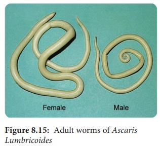
Adult worm
Ascaris lumbricoides resembles
and sometimes confused with the
earthworm. Its specific name lumbricoides
means earthworm in Latin. Male and Female worm of Ascaris lumbricoides are
shown in Figure 8.15.
The roundworm, Ascaris lumbricoides is the largest nematode parasite
in the human intestine. An editorialin the lancet in 189 observed, that if all
the round wor in all people worldwide were placed end -to-end. They would
encircle the worldtimes. Soil-transmitted intestinal nematodes are called Geohelminths.
• They
are large cylindrical worms with tapering ends. The anterior end being thinner
than the posterior end. It is the largest intestinal nematode parasitizing man.
• The
life – span of the adult worm is less than a year.
Male worm
• The
adult male worm is smaller than female worms .
• The
tail – end (Posterior end) of the male worm is curved ventrally to form a hook
and 2 curved copulatory spicules.
Female worm
• The adult female worm is larger (20–40 cm) and
thicker (3–6 mm) than male worm.
• The posterior end is conical and straight. The
anus is in the sub terminal part and opens like a transverse slit on the
ventral surface.
• The vulva is situated mid – ventrally, near the
junction of the anterior and middle thirds of the body. This part of the worm
is narrow and is called the vulvar waist.
• A single worm lays up to 200,000 eggs per day.
Egg: Two types of eggs are passed in feces by the worms.
Fertilized Egg
• The
fertilized eggs are produced by fertilized females.
• The
eggs are round or oval in shape and measures 45 µm in length and 35µm to 50µm
in breadth.
• They
are bile – stained and appear as golden brown (brownish) in colour.
• The egg
is surrounded by a thick smooth shell with an outer albuminous coat (corticated
eggs). Sometimes this outer coat is lost in few eggs. Those eggs are called as
decorticated eggs (Figure 8.16).
• Each
egg contains a large unsegmented ovum with a clear crescentic area at each
pole. The eggs float in saturated solution of common salt.
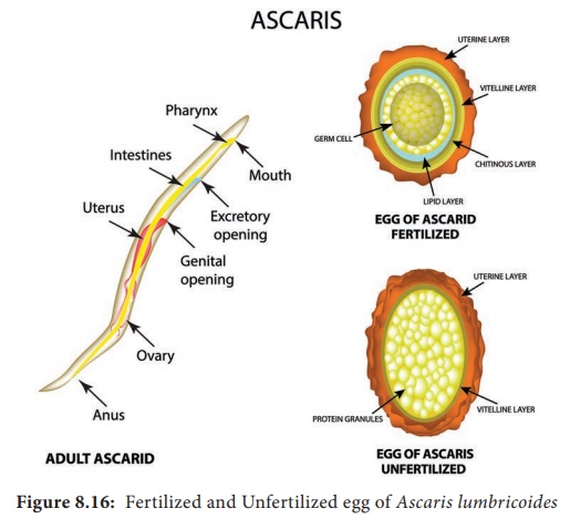
Unfertilized egg
• The female even not fertilized by male is capable
of liberating eggs. These unfertilized eggs are narrower, longer and elliptical
in shape.
• These are heaviest of all the helminthic eggs –
It measures about 80µm × 105µm in size.
• The eggs have a thinner shell with an irregular
coating of albumin (Figure 8.16).
• These
eggs do not float in saturated solution of common salt.
HOTS: What makes worm’s egg float or sink?
Life – Cycle
The life
– cycle of A. lumbricoides is
completed in a single host, human (Figure 8.17).
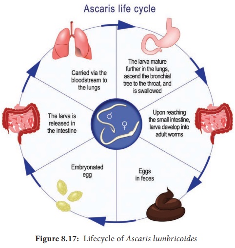
Infective form: Ermbryonated eggs. The fertilized egg passed in feces is not immediately
infective. It has to undergo a period of development in soil. The development
usually takes from 10–40 days. The embryo moults twice during the time and
becomes the infective rhabditiform larva.
Mode of transmission: Man
acquires the infection by ingestion
of food, water or raw vegetables contaminated with embryonated eggs of the
round worm.
The ingested eggs reach the duodenum to liberate the larvae by hatching. These larvae then penetrate the intestinal wall and are carried by the portal circulation to the liver. They live in liver for 3 to 4 days. Then they are carried to the right side of the heart, then to lung. In the lung, they grow and moult twice.
After
development in the lungs, in about 10–15 days, the larvae pierce the lung
capillaries and reach the alveoli. Then they are carried up the respiratory
passage to the throat and swallowed back to the small intestine.
In the
small intestine, the larvae moult finally and develop into adults. They become
sexually mature in about 6–12 weeks. The fertilized female start laying eggs
which are passed in the faces to repeat the cycle.
Pathogenesis
Infection
of A. lumbricoides in human is known
as ascariasis. The adult worm may produce its pathogenic effects in the
following ways.
a. The
spoliative or nutritional effects is usually seen when the worm burden is
heavy. Presence of enormous numbers (sometime exceeds 500) often interferes
with proper digestion and absorption of food. Ascariasis may contribute to
protein – energy malnutrition and vitamin A deficiency.
b. The
toxic effects is due to the metabolites of adult worm. Ascaris allergens
produce various allergic manifestations such as fever, urticaria and
conjunctivitis.
c. The mechanical
effects are the most important manifestations of ascariasis. In heavy
infections, adult worms can cause obstruction and inflammation of intestinal
tract, particularly of the terminal ileum.
d. Ectopic
ascariasis (Wander lust) is due to the adult male worms. They are restless wanderers.
Thewandering happens when the host temperature rises above 39°C. The worm may
wander up or down along the gut. It may enter the biliary or pancreatic duct
causing acute biliary obstruction or pancreatitis. It may enter the liver and
lead to liver abscesses. The worm may go up the esophagus and come out through
mouth or nose. It may crawl into the trachea and the lung causing respirator
obstruction or lung abscesses. Migrating downwards, the worm may cause
obstructive appendicitis. The worm may also reach kidneys. “Larva migrans” is a
term used when the larval sworms migrate to various parts of the body.
Clinical Manifestations
Incubation
Period is 60–70 days. Clinical manifestations due to adult worm vary from
asymptomatic to severe and even fatal infection. Clinical manifestation in
ascariasis can be caused either by the migrating larvae or by the adult worms.
Symptoms due to the migrating larvae: leads to
ascaris pneumonia and larvae may
enter the general circulation, disturbances have been reported in the brain,
spinal cord, heart and kidneys.
Symptoms due to the adult worms: Diffuse
or epigastric abdominal pain, abdominal cramping, abdominal swelling (especially
in children), fever, nausea, vomiting and passing roundworms and their eggs in
the stool.
Laboratory Diagnosis
Specimen
collected: Stool, sputum and blood.
Detection of parasite Adult worm: It can be
detected in stool or sputum of
patient by naked eye. Pancreatic or biliary worms can be detected by
ultra-sound and endoscope.
Larvae: Larvae can be detected in sputum and often in gastric washings. Chest X – ray may show
pulmonary infiltrates.
Eggs: Detection is through demonstration of eggs in feces. Detection of both fertilized and
unfertilized eggs are made after staining. Eggs may be demonstrative in the
bile obtained by duodenal aspirates.
Blood Examination
Complete
blood count may show eosinophilia in early stage of infection.
Serological tests
Ascaris
antibody can be detected by IHA, IFA and ELISA
Treatment
Commonly
used drugs are Albendazole and Mebendazole.
Prevention and Control
a. Proper
health education should be given for improved sanitation and personal hygiene.
b. Avoid eating of uncooked green vegetable, food
preparation and fruits that may contain faecal eggs.
c. Treating
infected persons especially children. Deworming of school children have been
found effective in control of ascariasis.
The National Deworming Day (Febuary 10th) is an (Febuary 10th)
is aninitiative of India to make every child in the country worm free. This is
one of the largest public health programs reaching large number of children
during a short period.
More than 836 million children are at risk of parasitic worm
infections worldwide. According to World Health Organization 241 million
children between the ages of 1 and 14years are at risk of parasitic intestinal
worms in India, also known as Soil-Transmitted Helminths (STH).
Related Topics