Chapter: Microbiology and Immunology: Bacteriology: Staphylococcus
Laboratory diagnosis of staphylococcal infections - Staphylococcus aureus
Laboratory Diagnosis
Laboratory diagnosis of staphylococcal infections is based on the demonstration of staphylococci, in appropriate clinical specimens, by microscopy and culture.
◗ Specimens
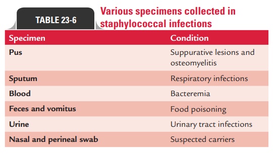
Specimens to be collected for demonstration of staphylococci depend on the nature of lesion (Table 23-6).
◗ Microscopy
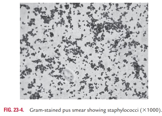
Demonstration of Gram-positive cocci arranged in clusters and pus cells in the Gram-stained smears of pus (Fig. 23-4, Color Photo 14), wound exudate, etc. are the characteristic features of pyogenic infection caused by S. aureus. It is noteworthy that microscopy:
· Alone is not adequate to differentiate various species of staphylococci or micrococci from one another.
· Is also of no value for sputum and other specimens where mixed bacterial flora is present.
◗ Culture
The identification of staphylococci is confirmed by culture and other identification tests comprising a range of biochemical and enzymatic tests followed by antibiotic sensitivity. The spec-imens are inoculated onto nutrient agar and blood agar and incubated at 37°C for 24 hours. On nutrient agar, large, circu-lar, smooth, convex, and glistening colonies showing golden-yellow pigments can be observed. On blood agar, the colonies show a zone of beta-hemolysis, which is not shown by any other species of staphylococci.
Specimens from heavily contaminated sources, such as vom-itus and feces, are inoculated on selective media (e.g., manni-tol salt agar or salt milk agar). These media inhibit growth of Gram-negative bacteria but allow the growth of staphylococci and certain other Gram-positive cocci.
◗ Identification of bacteria
The identifying features of S. aureus are summarized.
Coagulase test
Coagulase test is an important test carried out to detect S.aureus. The test is done in two ways: tube coagulase test andslide coagulase test.
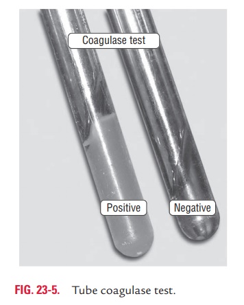
Tube coagulase test: Tube coagulase test is carried out todetect free coagulase. In this test, 0.1 mL of an overnight broth culture is mixed with 0.5 mL of a 1:10 dilution of human or rabbit plasma. The plasma-broth culture mixture is incu-bated in a water bath at 37°C for 3–6 hours. In a positive test, the plasma is coagulated and does not flow (Fig. 23-5, Color Photo 13).
Human or rabbit plasma, which is rich in CRF, is used in the test. The plasma is collected in vials containing anticoagu-lants, such as oxalate, heparin, or EDTA.
Citrated plasma is not used because if the specimen is con-taminated with Gram-negative bacilli, the latter may utilize the citrate and produce false positive reaction.
Slide coagulase test: Slide coagulase test detects the boundcoagulase or the clumping factor. The test is performed by mix-ing a dense suspension of the staphylococci with a loopful of undiluted rabbit plasma on a slide. In a positive test, clumping takes place within 10 seconds.
Phosphatase test
The production of phosphatase can be demonstrated by culturing a mixed specimen on phenolphthalein phosphate agar and exposing the colonies to ammonium vapors. S. aureus colonies turn bright pink due to the release of phenolphthalein.
Novobiocin sensitivity
Novobiocin sensitivity is a simple disk diffusion test to differ-entiate S. aureus from other staphylococci. This test is carried out by using a 5-mg novobiocin disk on an overnight culture of staphylococci on Mueller–Hinton agar. Novobiocin sensitivity is shown by an inhibition zone of $16 mm. S. aureus is novobio-cin sensitive, while S. saprophyticus is novobiocin resistant.
Polymyxin B resistance
Polymyxin B sensitivity is again a simple disk diffusion test to differentiate S. aureus from other staphylococci. This is carried out by using a 300-U polymyxin B disk on an overnight culture of staphylococci on Mueller–Hinton agar. Polymyxin resistance is shown by an inhibition zone of ,10 mm. S. aureus is usually polymyxin resistant.
Related Topics