Chapter: Introduction to Human Nutrition: Body Composition
Imaging techniques - Indirect methods in Body composition techniques
Imaging techniques
CT scanning enables the visualization of tissues in cross-sectional slices of the body. The thickness of those slices can vary, but is normally about 1 cm. During CT scanning a source of X-rays rotates per-pendicularly around the body or a body segment, while photodetectors, opposite to the source, register the attenuation of the X-rays after they have passed through the body in the various directions. The infor-mation received by the photodetectors is used to gen-erate images. Software enables the calculation of the

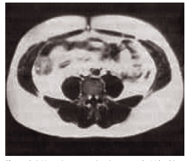
Figure 2.4 Magnetic resonance imaging scan at the L4 level in an obese subject. The white areas in the image are adipose tissue. Sub-cutaneous adipose tissue and intra-abdominal adipose tissue are sepa-rated by the abdominal muscles.
amounts of tissues with different attenuation, for example adipose tissue against nonadipose tissue. The CT technique was introduced for body composi-tion assessments in the 1980s and is now widely used, predominantly for measurements of body fat distri-bution. Figure 2.4 shows a scan of the abdomen at the level of the umbilicus, made by MRI, a technique that gives comparable information. The precision of the calculation of a tissue area or tissue volume from the same scan(s) is very accurate, with an error of about 1%. Partial volume effects (pixels that contain tissue with different attenuation) may influence the accu-racy and reproducibility of the method.
A single CT scan provides only relative data, for example in a scan of the abdomen the relative amount of visceral adipose tissue to subcutaneous adipose tissue. Multiple CT scanning allows the calculation of tissue volumes. From adipose tissue volumes (tissue level) and an assumed density and composition of the adipose tissue, the amount of fat mass (molecular level) can be calculated. Multiplying tissue volumes with specific densities of these tissues (determined in vitro) allows a recalculation of the body weight, a necessary but not sufficient exercise for validation of a whole body technique. Research in this area has shown that the CT technique allows the determina-tion of total body composition, with an error of estimate for fat mass of 3–3.5 kg (compared with densitometry).
CT scanning is expensive and, because of the rela-tively high level of radiation, the method is limited to subjects for whom scanning is indicated on clinical grounds. An alternative method to CT scanning is MRI, which has the advantage that no ionizing radia-tion is involved.
During MRI, the signals emitted when the body is placed in a strong magnetic field are collected and, as with CT scanning, the data are used to generate a visual cross-sectional slice of the body in a certain region. The determination of adipose tissue versus nonadipose tissue is based on the shorter relaxation time of adipose tissue than of other tissues that contain more protons or differ in resonance frequency. MRI has the advantage over CT scanning that the subject is not exposed to ionizing radiation. However, the time necessary to make an MRI image is relatively long (minutes versus seconds using CT), which has impli-cations for the quality of the image. Any movement of the subject, even the movements of the intestinal tract when making images in the abdominal region, will decrease the quality of the image.
As with CT scanning, images can be combined to obtain information on total body composition. Infor-mation about organ size can be obtained with a high accuracy. For example, MRI is used to study the con-tribution of various organs to the resting metabolic rate of the total body.
Both CT scanning and MRI are expensive, and therefore their use will remain limited to a few labo-ratories and for very specific situations.
Related Topics