Structure, Classification - Herpes Viruses | 12th Microbiology : Chapter 10 : Medical Virology
Chapter: 12th Microbiology : Chapter 10 : Medical Virology
Herpes Viruses
Herpes Viruses
The
herpes virus family contains more than a hundred species of enveloped DNA viruses that affect
humans and animals.
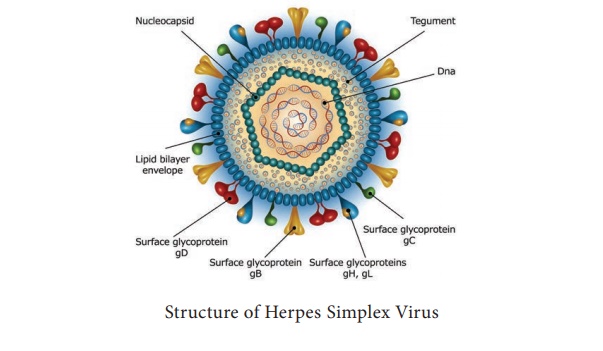
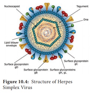
Structure
The
herpes virus capsid is icosahedral,
composed of 162 capsomers and enclosing the core containing the linear double stranded DNA genome. The nucleocapsid is surrounded by the lipid envelope derived from the host cell.
The envelope carries surface spikes (Figure
10.4). Teguments are present in
between the envelope and capsid. The enveloped
virion measures about 200nm and the naked
virion about 100 nm in diameter.
Classification
Herpes
virus belongs to the family Herpesviridae.
i. Alpha herpes viruses
They have
relatively short replicative cycle (12–18 hours) and a variable host range. They cause latent infection in
sensory ganglia. Example: Herpes simplex virus and varicella zoster virus.
ii. Beta herpes viruses
They replicate slowly (more than 24 hours) and have a narrow host
range, grow well in fibroblasts. They cause latent infection of salivary gland
and other organs. Example: Cytomegalovirus.
iii. Gamma herpes viruses
They have
a narrow host range and replicate in
lymphoblastoid cells. They are specific for either B or T lymphocytes and
causes latent infection in lymphoid tissue Example: Epstein - Barr Virus.
Eight
different types of herpes viruses are known whose primary hosts are humans.
They have been designated as ‘Human herpes virus type 1–8.
1. Herpes Simplex
The
herpes simplex virus (HSV) occurs naturally
only in humans, but it can produce
experimental infection in laboratory animals. There are two types of the herpes simplex virus. HSV type 1 (Human herpes virus type 1) is isolated from lesions in
and around the mouth and is transmitted by direct contact or droplet spread
from carrier. HSV type 2 (Human
herpes virus type 2 or HHV type 2)
is responsible for the genital herpes infections
transmitted venereally.
Pathogenesis
Herpes
simplex is one of the most common viral
infection in humans, the sources
of infection are saliva, skin lesions or respiratory secretions. In type 2,
transmission occurs by close contact and may be veneral in genital herpes.
The virus
enters through defects in the skin or mucous membranes and multiples locally,
with cell to cell spread. The herpes
lesions are thin walled, umbilicated vesicles, the roof of which breaks
down, leaving tiny superficial ulcers. They heal without scarring.
Clinical features
The
clinical manifestations depend on the site of infection, age and immune status
of the host and the antigenic type of the virus. They are
• Cutaneous
infections
• Mucosal
infections
• Ophthalmic
infections
• Nervous
system infections
• Visceral
infections
• Genital
infections
Laboratory diagnosis
Microscopy
Smears
are prepared from the lesions, from the vesicles and stained with 1% aqueous
solution of toluidine blue ‘O’ for
15 seconds. Multinucleated giant cells with faceted nuclei with ground glass
chromatin (Tzanck cells) are
observed.
Virus isolation
Inoculation
in mice and on chick embryo CAM is insensitive. Primary human embryonic kidney,
human amnion cells are susceptible, but human
diploid fibroblasts are
preferred. Vesicle fluid, spinal
fluid, saliva and swabs may be used. Cytopathic changes may appear as early as
24–48 hrs.
Serology
Antibodies
develop within a few days of infection and rise
in titre of antibodies may be demonstrated by ELISA, neutralization or
complement fixation tests.
Chemotherapy
Indoxyuridine
used topically in eye and skin infection, acyclovir and vidarabine are given
for deep and systemic infections.
2. Varicella Zoster
In 1889, Von Bokay had
suggested that varicella (Chicken
pox) and herpes zoster are different
manifestations of the same virus infection. The virus is therefore called Varicella zoster virus (VZV). The
chicken pox follows primary infection
in a non immune individual, while herpes zoster is a reaction of the latent virus when the immunity has fallen to
infective levels.
VZV is similar to the herpes simplex virus in
its morphology. It can be grown in cultures of human fibroblasts human amnion
or HeLa cells. Chicken pox is one of
the mildest and most common of child
hood infections. The disease may,
occur at any age.
Herpes gladiatorum is spread through
skin-to-skin contact. Ifyoukiss someone with a herpes cold sore on their lips, you
could become infected.
3. Cytomegaloviruses
Cytomegaloviruses
(CMV) formerly known as salivary gland
viruses are a group of ubiquitous herpes viruses of humans and animals.
They are characterized by enlargement of
infected cells and intranuclear
inclusions. In 1926, cytomegalia
presumed to be due to viral infection was reported in the salivary glands of
guinea pigs and children and the viral agent was called the ‘salivary gland virus’
Infobits: CMV is the largest
viruses in the herpes virus family, being 150–200 nm in size.
4. Epstein – Barr Virus
A number
of different viruses apparently ‘Passenger
Viruses’ were isolated from cultured
lymphoma cells. Epstein, Barr and Achong in 1964 observed a new type of herpes virus and named it has ‘EB Virus’ affecting B lymphocytes of only human and some sub human primate B cells have receptors ( CD21 molecules) for the virus.
The
source of infection is usually the saliva
of infected persons who shed the
virus in oropharyngeal secretions. Intimate oral contact, as in kissing,
appears to be the predominant mode of transmission. This accounts for
infectious mononucleosis being called as ‘The
kissing disease’.
5. Human Herpes Virus Types 6,7,8
A herpes
virus, first isolated in 1986 from the peripheral
blood of patients with lympho proliforative disease called as human B lymphotropic virus, renamed as HHV - 6. HHV- 7 was isolated in
1990 from peripheral CD4 cells of a
healthy person appears to be widely distributed and transmitted through saliva.
In 1994,
DNA sequences presumed to represent a new herpes virus from tissues of Kaposi’s sarcoma from AIDS patients was named as HHV8. Later Kaposi’s sarcoma was identified in persons
not infected with HIV and referred to as Kaposi’s
Sarcoma-associated Herpes Virus (KSHV)
Related Topics