Chapter: Ophthalmology: Eye Optics and Refractive Errors
Eye Optics and Refractive Errors: Examination Methods
Examination Methods
Refraction Testing
Refraction testing means measuring the additional refractive power required to
produce a sharp image on the retina. Subjective and objective methods are used.
Subjective methods require information from the patient.
Subjective refraction testing:
This consists of successively placing variouscombinations of
lenses before the patient’s eye until the maximum visual acuity is reached (see
Correction of Refractive Errors).
Objective refraction testing:
Objective testing is unavoidable when thepatient is unable to
provide subjective information (for example with infants) or when this
information is unreliable. This method also greatly accelerates subjective
refractive testing.
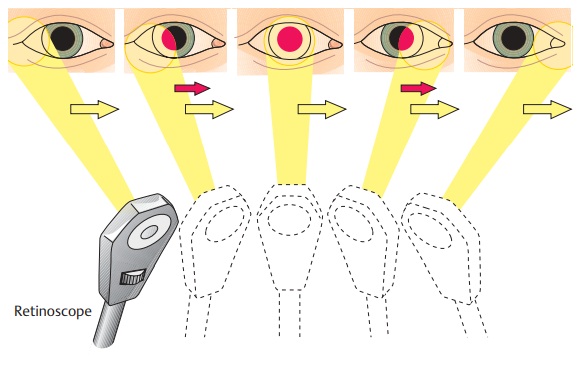
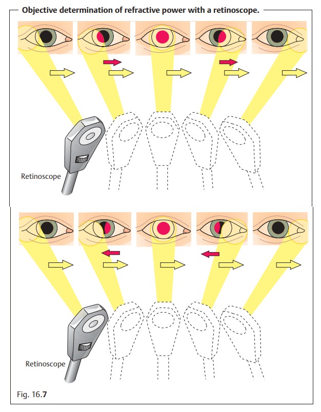
Retinoscopy (shadow testing): The retina is illuminated through the pupil.The examiner observes the optical phenomena in the patient’s pupil while moving the light source (Fig. 16.7).
Refractometry.The measuring principle is based on ophthalmoscopic obser-vation
of a test image projected on to the patient’s retina. The distance between the
test figure and the eye is changed until the image appears in focus on the
retina. Refraction can then be calculated from the measured values. An
alternative to changing of the distance is to place various lenses in the path
of the light beam.
Automated refractometry.The method measures refraction automaticallywith the aid of
light-sensitive detectors and a computer until a focused image appears on the
retina. These systems operate with infrared light.
Any objective measurements of refraction
should be verified by subjec-tive testing whenever possible.
Testing the Potential Resolving Power of the Retina in the Presence of Opacified Ocular Media
Special examination methods are indicated in
the presence of opacification of the ocular media of the eye (such as a
cataract) to determine the potential visual acuity of the retina. This permits
the ophthalmologist to estimate whether optimizing the refractive media with
techniques such as cataract surgery or corneal transplantation would achieve
the desired improvement.
Laser interference visual acuity testing:
Lasers are used to project inferencestrips of varying widths on to the retina. The patient must specify the direc-tion in which these increasing narrower strips are aligned. This examination can no longer be performed where there is severe opacification of the optic media such as in a mature cataract. The preliminary examination then con-sists of evaluating the pattern of the transilluminated retinal vasculature.
Fig. 16.7 With the
retinoscope, the examiner moves a light source (a beam of yellow light) across
the pupil (dark spot) at a distance of about 50 cm from the patient. This
pro-duces a light reflex (red spot) in the patient’s eye. It is important to
note how this light re-flex (red spot) behaves as the light source of the
retinoscope is moved. There are two possibilities:
a “With” motion: the light reflex in the pupil (red spot) moves in the same
direction (redarrows) as the light source of the retinoscope (yellow arrows).
This means that the far point of the eye is behind
the light source. b “Against” motion.
The light reflex in the pupil moves in the opposite
direction (red arrows) to the light source of the retinoscope (yellow arrows).
This means that the far point of the eye lies between the eye and the light source. The examiner places
appropriate lenses in front of the patient’s eyes (plus lenses for “with”
motion and minus lenses for “against” motion) until no further motion of the
light reflex is observed. The motion of the retinoscope will then only elicit an
infinitely fast reflex (neutral point).
This method is used to determine the proper lens for correct-ing the refractive
error.
Related Topics