Chapter: BIOLOGY (ZOOLOGY) Standard XI first year 11th text book Assignment topics question and answer Explanation Definition
Endoplasmic Reticulum (ER) and Function of Endoplasmic Reticulum

Endoplasmic Reticulum. (ER)
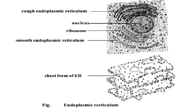
Electron microscopic study of sectioned cells has revealed the pres-ence of a three dimensional network of sac-like and tubular cavities calledcisternae bounded by a unit membrane inside the cell. Since these structures are concentrated in the endoplasmic portion of the cytoplasm, the entire organisation is called the endoplasmic reticulum. This name was coined by Porter in 1953.
The occurrence of ER varies from cell to cell. They are absent in erythrocytes, egg cells and embryonic cells.
The ER is the site of specific enzyme controlled biochemical reactions. Its outer surface carries numerous ribosomes. The presence of ribosomes gives a granular appearance . In this condition ER is described as rough endoplasmic reticulum (RER). RER is the site of syn-thesis of proteins. Ribosomes are absent on smooth endoplasmic reticulum (SER). SER is concerned with lipid metabolism.
Morphologically ER may occur in three forms namely
1. Lamellar form
2. Vesicular form and
3. Tubular form.
Lamellar form or Cisternae :- These are long, flat, sac like tubules. Their diameter is about 40-50 Am. The RER has a synthetic role. It is mostly seen in cells of pancreas, notochord and brain.
Vesicles :- These are oval, vacuolar structures. Their diameter is about 25-500 Am. They occur in most of the cells.
Tubules :- These are branched structures forming the reticular system along with the cisternae and vesicles. They have a diameter of 50-190 Am. They occur in almost all cells.
Functions :-
1. It provides skeletal framework to the cell.
2. It facilitates exchange of molecules by the process of osmosis, diffusion and active transport.
3. Enzymes of ER control several metabolic activities.
4. They serve as intracellular transporting system.
5. It conducts intra-cellular impulses.
6. It helps to form nuclear membrane after cell division.
Related Topics