Chapter: Obstetrics and Gynecology: Embryology and Anatomy
Embryology and Anatomy

Embryology and Anatomy
Knowledge of the embryology and anatomy
of the female genital system is helpful in understandingboth normal
anatomy and the congenital anom-alies that occur. Embryology may be useful in
many areas of gynecologic and obstetric practice. For example,
in gynecologic oncology, embryology can assist clinicians in predicting the
growth and routes of spread of gynecologic cancers; in urogynecology and pelvic
reconstructive surgery, it can enhance a surgeon’s comprehension of the
compo-nents of pelvic support and possible defects. It can also play a key role
in understanding and diagnosing various aspects of sexual dysfunction.
The ovaries, fallopian tubes,
uterus, and upper portion of the vagina are derived from the intermediate
mesoderm, while the external genitalia develop from genital swellings in the
pelvic region. Beginning in the 4th week (postfertil-ization) of development,
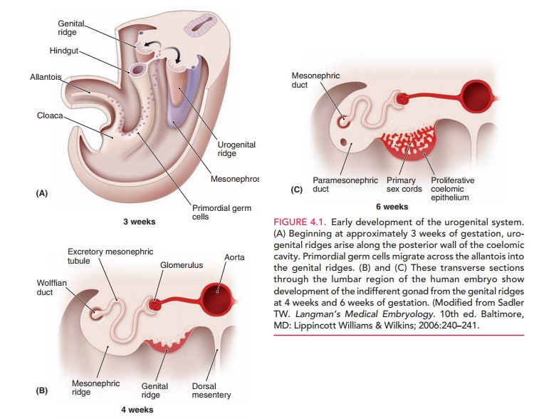
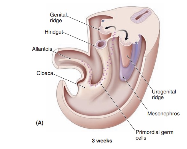
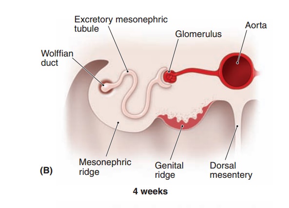
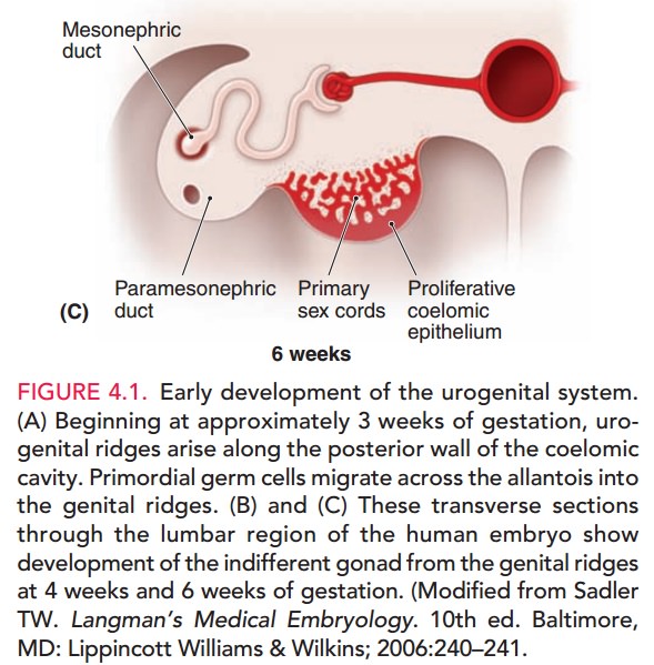
the intermediate mesoderm forms the urogenital
ridges along the posterior body wall. As their name implies, these ridges
contribute to the forma-tion of the urinary and genital systems (Fig. 4.1).


The
gonads, genital ducts, and external genitalia all pass through an indifferent (undifferentiated) stage in
which it is not possible to determine sex based on the appearance of these
struc-tures. The genetic sex of an embryo is determined by the
sexchromosome (X or Y) carried by the sperm that fertilizes the oocyte. The Y
chromosome contains a gene called SRY (sex-determining region on Y) that encodes a protein called testis-determining factor (TDF). When
this pro-tein is present, the embryo develops male sex characteris-tics. The
ovary-determining gene is WNT4; when this gene is present and SRY is absent, the embryo develops
female characteristics. Gonads become
structurally male or female bythe 7th week of development, and external
genitalia become dif-ferentiated by the 12th week. The influence of
androgens iscrucial in the normal development of the external geni-talia. Any
condition that increases the level of androgen production in a female embryo
will cause developmen-tal anomalies. For example, the genetic disease congen-ital adrenal hyperplasia (CAH) causes
a decreasedproduction of cortisol that results in a compensatory increase in
androgens. The genitalia of female fetuses with CAH are ambiguous, that is,
neither normal female nor normal male.
Related Topics