Chapter: Obstetrics and Gynecology: Embryology and Anatomy
Development of the External Genitalia
Development of the External
Genitalia
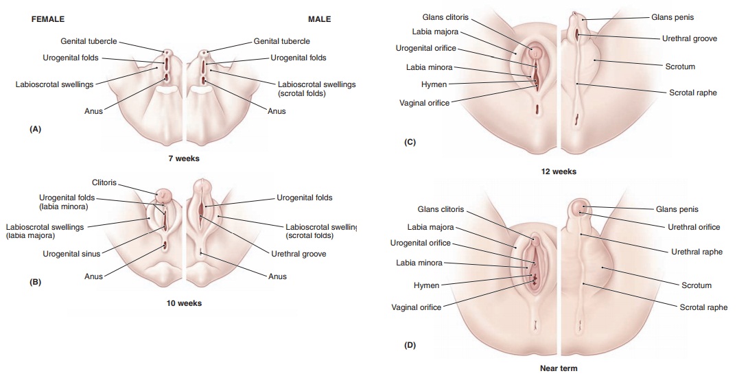
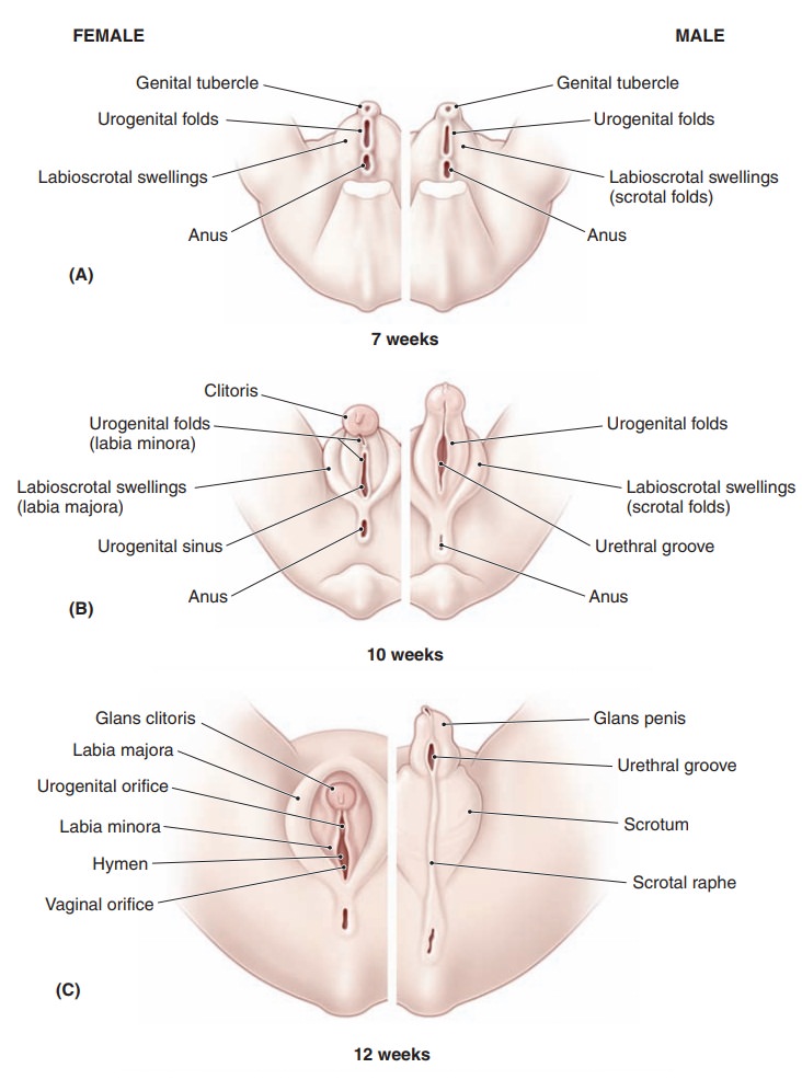
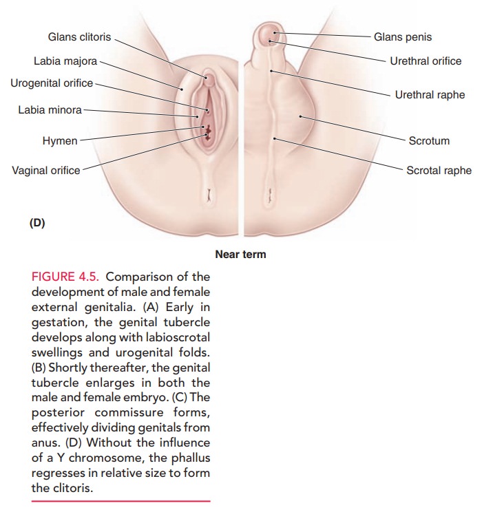
The cloaca is formed from a dilatation of the caudal end of the hindgut and is covered exteriorly by the cloacal mem-brane. Eventually, the cloaca is separated into the urogeni-tal sinus anteriorly and the anorectal canal posteriorly by the urorectal septum. This septum forms from a collec-tion of mesoderm in the pelvic floor that grows downward during the 5th to the 8th weeks of gestation to reach the cloacal membrane. At the same time, the genital tuber-cle develops at the cranial end of the cloacal membrane,while labioscrotal swellings and urogenital folds appear on each side (Fig. 4.5A). The genital tubercle enlarges in both the male and the female (Fig. 4.5B). In the presence of estrogens and the absence of androgens, external genitalia are feminized. The genital tubercle develops into the clitoris (Fig. 4.5C). The unfused urogenital folds form the labia minora, and the labioscrotal swellings become the labia majora (Fig. 4.5D).
At
approximately 15 weeks of gestation, transverse ultra-sonography can
distinguish between the two sexes, although it is not definitive.
Related Topics