Chapter: Essentials of Anatomy and Physiology: The Urinary System
Elimination of Urine
ELIMINATION OF URINE
The ureters, urinary bladder, and urethra do not change the composition or amount of urine, but are responsible for the periodic elimination of urine.
URETERS
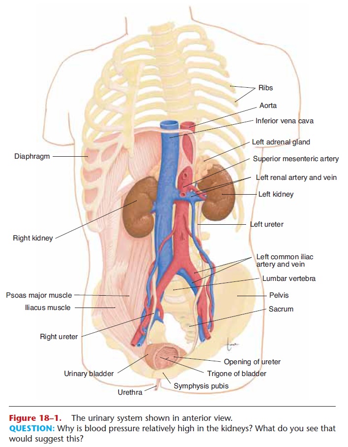
Each ureter extends from the hilus of a kidney to the lower, posterior side of the urinary bladder (see Fig. 18–1). Like the kidneys, the ureters are retroperi-toneal, that is, behind the peritoneum of the dorsal abdominal cavity.
The smooth muscle in the wall of the ureter con-tracts in peristaltic waves to propel urine toward the urinary bladder. As the bladder fills, it expands and compresses the lower ends of the ureters to prevent backflow of urine.
URINARY BLADDER
The urinary bladder is a muscular sac below the peri-toneum and behind the pubic bones. In women, the bladder is inferior to the uterus; in men, the bladder is superior to the prostate gland. The bladder is a reser-voir for accumulating urine, and it contracts to elimi-nate urine.
The mucosa of the bladder is transitional epithe-lium, which permits expansion without tearing the lining. When the bladder is empty, the mucosa appears wrinkled; these folds are rugae, which also permit expansion. On the floor of the bladder is a tri- angular area called the trigone, which has no rugae and does not expand. The points of the triangle are the openings of the two ureters and that of the urethra (Fig. 18–7).
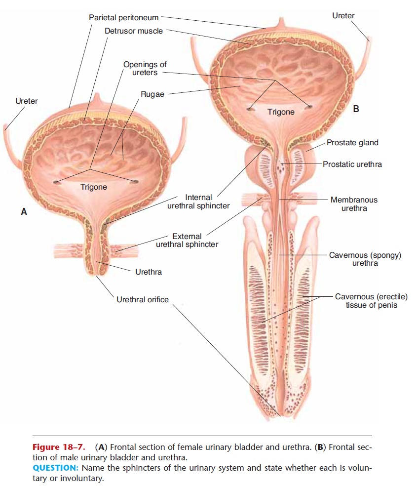
Figure 18–7. (A) Frontal section of female urinary bladder and urethra. (B) Frontal sec-tion of male urinary bladder and urethra.
QUESTION: Name the sphincters of the urinary system and state whether each is volun-tary or involuntary.
The smooth muscle layer in the wall of the bladder is called the detrusor muscle. It is a muscle in the form of a sphere; when it contracts it becomes a smaller sphere, and its volume diminishes. Around the opening of the urethra the muscle fibers of the detru-sor form the internal urethral sphincter (or sphinc-ter of the bladder), which is involuntary.
URETHRA
The urethra (see Fig. 18–7) carries urine from the bladder to the exterior. The external urethral sphincter is made of the surrounding skeletal muscle of the pelvic floor, and is under voluntary control.
In women, the urethra is 1 to 1.5 inches (2.5 to 4 cm) long and is anterior to the vagina. In men, the urethra is 7 to 8 inches (17 to 20 cm) long. The first part just outside the bladder is called the prostatic ure-thra because it is surrounded by the prostate gland. The next inch is the membranous urethra, around which is the external urethral sphincter. The longest portion is the cavernous urethra (or spongy or penile urethra), which passes through the cavernous (or erec-tile) tissue of the penis. The male urethra carries semen as well as urine.
THE URINATION REFLEX
Urination may also be called micturition or voiding. This reflex is a spinal cord reflex over which voluntary control may be exerted. The stimulus for the reflex is stretching of the detrusor muscle of the bladder. The bladder can hold as much as 800 mL of urine, or even more, but the reflex is activated long before the maxi-mum is reached.
When urine volume reaches 200 to 400 mL, the stretching is sufficient to generate sensory impulses that travel to the sacral spinal cord. Motor impulses return along parasympathetic nerves to the detrusor muscle, causing contraction. At the same time, the internal urethral sphincter relaxes. If the external ure-thral sphincter is voluntarily relaxed, urine flows into the urethra, and the bladder is emptied.
Urination can be prevented by voluntary contrac-tion of the external urethral sphincter. However, if the bladder continues to fill and be stretched, voluntary control is eventually no longer possible.
Related Topics