Reproductive Health - Detection of foetal disorders during early pregnancy | 12th Zoology : Chapter 3 : Reproductive Health
Chapter: 12th Zoology : Chapter 3 : Reproductive Health
Detection of foetal disorders during early pregnancy
Detection
of foetal disorders during early pregnancy
Ultrasound scanning
Ultrasound has no known
risks other than mild discomfort due to pressure from the transducer on the
abdomen or vagina. No radiation is used during this procedure. Ultrasonography
is usually performed in the first trimester for dating, determination of the
number of foetuses, and for assessment of early pregnancy complications.
There are several
types of ultrasound imaging techniques. As the most common type, the 2-D
ultrasound provides a flat picture of one aspect of the baby. The 3-D image
allows the health care provider to see the width, height and depth of the
images, which can be helpful during the diagnosis. The latest technology is 4-D
ultrasound, which allows the health care provider to visualize the unborn baby
moving in real time with a three-dimensional image.
Amniocentesis
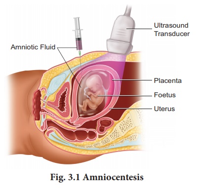
Amniocentesis involves
taking a small sample of the amniotic fluid that surrounds the foetus to diagnose
for chromosomal abnormalities (Fig. 3.1).

Amniocentesis is
generally performed in a pregnant woman between the 15th and 20th weeks of
pregnancy by inserting a long, thin needle through the abdomen into the
amniotic sac to withdraw a small sample of amniotic fluid. The amniotic fluid
contains cells shed from the foetus.
Chorionic villus sampling (CVS)
CVS is a prenatal test
that involves taking a sample of the placental tissue to test for chromosomal
abnormalities.
Foetoscope
Foetoscope is used to
monitor the foetal heart rate and other functions during late pregnancy and
labour. The average foetal heart rate is between 120 and 160 beats per minute.
An abnormal foetal heart rate or pattern may mean that the foetus is not getting
enough oxygen and it indicates other problems.
A hand-held doppler
device is often used during prenatal visits to count the foetal heart rate.
During labour, continuous electronic foetal monitoring is often used.
BREAST SELF EXAMINATION AND EARLY DIAGNOSIS OF CANCER
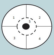
1. Breast is divided into 4
quadrants and the center (Nipple) which is the 5th quadrant.
2. Each quadrant of the breast is felt
for lumps using the palm of the opposite hand.
3. The examination is done in both
lying down and standing positions, monthly once after the 1st week of menstrual
cycle.
This way if there are lumps or any
deviation of the nipple to one side or any blood discharge from the nipple we
can identify cancer at an early stage.

Mammograms are done for women above
the age of 40 years and for young girls and women below 40 years. Ultrasound of
the breast aids in early diagnosis.
• Vitamin E is known as
anti-sterility vitamin as it helps in the normal functioning of reproductive
structures.
• Sex hormones were discovered by
Adolf Butenandt.
• 11th July is observed as World
Population Day.
• 1st December is observed as World
AIDS Day.
• NACO (National AIDS Control
Organisation) was established in 1992.
• Syphilis and gonorrhoea are
commonly called as international diseases.
Related Topics