Chapter: BIOLOGY (ZOOLOGY) Standard XI first year 11th text book Assignment topics question and answer Explanation Definition
Components of Human Circulatory system : Blood vessels, Types of blood vessels, The Heart
Components of Human Circulatory system : Blood vessels, Types of blood vessels, The Heart
Components of Circulatory system
Blood vessels
The blood vessels carrying blood away from the heart are the arteries. The Veins carry blood towards the heart. The arteries and veins are named and classified according to their anatomical position. They can also be classified according to their size and wall structure. Functionally, arteries are subdivided into conducting, distributing and resistance vessels.
1. Conducting vessels :- These are large arteries from the heart and their main branches. the walls of these vessels are elastic in nature.
2. Distributing vessels :- These are smaller arteries reaching individual organs. They branch into the organs. They have muscular walls.
3. Resistance vessels :- These are mostly arterioles. While these vessels are smaller, their walls are highly muscular. Hence these vessels can reduce pressure of blood due to peripheral resistance.
4. Exchange vessels :- These are the capillaries. The walls of these vessels allow exchanges between blood and the tissue fluid surrounding the cells. The substances commonly exchanged are oxygen, carbon-di-oxide, nutrients, water, inorganic ions, vitamins, hormones, metabolic products and antibodies.
5. Capacitance or reservoir vessels :- These are the larger vessels and veins. These are of varying sizes. They collect and convey blood back to the heart. The higher capacitance of these vessels is due to their distensibility. Hence their blood content is more, even at low pressure. The number of such veins is also enormous.. Thus the veins are called as the ' blood reservoirs'
Structure of blood vessels
The blood vessels show a vast range of structural modifications. However a few basic patterns can be studied.
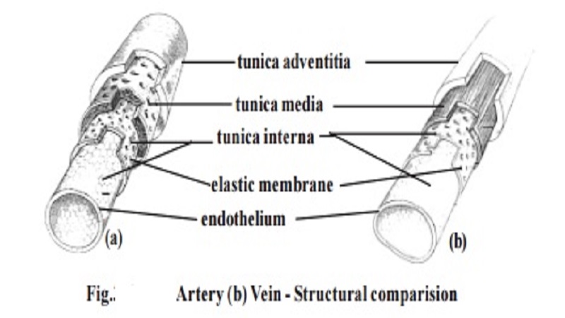
A blood vessel consists of a wall and a lumen or cavity. The wall of the blood vessels is made up of 3 distinct layers or tunica. They are the tunica intima, tunica media and tunica externa or tunica adventitia.
The tunica intima is formed of an endothelium, a delicate connective tissue and elastic fibres. The tunica media contains smooth muscle cells. It causes vasoconstriction and vasodilation. The tunica externa is composed of connective tissue. The composition and thickness of layers varies with the diameter of the blood vessels and the type.
Types of blood vessels
1. Large elastic arteries :- The walls of these arteries contain elastic fi-bres. The smooth wall measures about 1micron in thickness. It gets stretched under the effect of pulse and recoils elastically.
2. Muscular arteries :- There are larger and smaller muscular arteries. The larger muscular arteries are inelastic and they have thick walls. The wall has 30-40microns in diameter in the layers of smooth muscles. Since they regulate blood supply, they are called distributing arteries. The small muscular arteries are capable of vasodilation and vasoconstriction.
3. Arterioles :- They conduct blood from the arteries to the capillary bed. These are small vessels capable of vasodilation and vasoconstriction.
4. Capillaries :- These are fine vessels found between arterioles and venules. They measure 5-8micron in diameter.
5. Venules :- These are tubes of flat, oval or polygonal endothelial cells. Each venule is formed by the convergence of two or more capillaries. Its diameter ranges upto 30micron.
6. Veins :- Veins seen in anatomy are medium veins. They run in between venules and large veins. Large veins transport blood to the heart.
Veins with diameter above 2 mm have valves. They are of semilunar type. They allow movement of blood towards the heart. There are several valves in the medium veins.
Branching of blood vessels :- When an artery divides into two equal branches, the original artery ceases to exist. Hence the branches are called terminalbranches. The smaller branching vessels formed on the sides are called the collateral branches. When arteries are joined to each other it is named asanastomosis.
Blood supply to blood vessels :- As any other region, the cells and tissue on the wall of the blood vessel require nourishment. Some amount can diffuse from blood in the lumen. For vessels having diameter greater than 1 mm, dif-fusion of nutrients may not be possible. Such vessels have very minute ves-sels called vasa vasorum spread over them. They penetrate into the wall of the blood vessels.
Innervation of blood vessels :- The walls of the blood vessels are inner-vated by sympathetic nerve fibres. They regulate the contraction of the mus-culature. They effect vasoconstriction.
The Heart
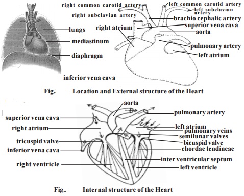
The heart is a hollow, fibromuscular organ. It is somewhat conical or pyramidal in form. It is roughly the size of a closed fist. An average heart measures 12 cm from base to the apex. Transverse diameter at its broadest region is 8-9 cm. It is 6 cm thick antero-posteriorly. While in adult male the heart weighs 280-340 g, in female it weighs 230-280 g.
The thoracic organs such as heart, trachea and oesophagus form a midline partition called the mediastinum. The heart lies obliquely in the medi-astinum.
The heart is surrounded by a double layered membrane called the peri-cardium. The outer layer is called the fibrous pericardium. The inner mem-brane is called the serous pericardium. In between heart and pericardium, there is a pericardial space. This space is filled with a fluid called the pericardial fluid.
The wall of the heart is made up of three tissue layers. They are the epicardium, myocardium and endocardium. The epicardium forms the smooth outer surface of the heart. The middle myocardium is composed of cardiac muscle. This layer plays an important role in the functioning of the heart. Theendocardium forms the smooth inner surface. It is formed of squamous epithelium.
Related Topics