Chapter: Obstetrics and Gynecology: Assessment of Genetic Disorders in Obstetrics and Gynecology
Chromosome Replication and Cell Division
Chromosomes
The genetic information in the
human genome is packaged as chromatin,
within which DNA binds with several chromosomal proteins to make chromosomes. A karyo-type reveals the morphology and number of chromo-somes. Somatic cells are all the cells in the
human body that are not gametes (eggs or sperm). Germ cells, or gametes, contain a single set of chromosomes (n= 23) and are described as haploid in number. Somatic cells
contain two sets of chromosomes, for
a total of 46 chromosomes.These cells are diploid,
signifying that they have a 2n chro-mosome complement (2n = 46). These chromosome pairs
consist of 22 pairs of autosomes,
which are similar in males and females. Each somatic cell also contains a pair
of sex chromosomes. Females have two X sex chromosomes; males have an X and a Y
chromosome.
CHROMOSOME REPLICATION AND CELL DIVISION
Chromosomes undergo two types of replication, meiosis and mitosis, which
are significantly different and produce cell types with crucially different
capabilities. Mitosis is the
replication of chromosomes in somatic cells. It is fol-lowed by cytokinesis, or cell division, that
results in two daughter cells containing the same genetic information as the
parent cell. Meiosis only occurs in
germ cells. It is also followed by cytokinesis; but, in this case, cytokinesis
results in four daughter cells with a haploid count.
Somatic cells undergo cell
division based on the cell cycle. There are four stages of the cell cycle: G1,
S, G2, and M. G1, or gap 1, occurs immediately after mitosis and is a period of
inactivity with no DNA replication. During G1
all the DNA of each chromosome is present in the 2n form. The next phase is S, or synthesis, where the chromosomes
double to become two identical sister chromatids with a 4n chromosome
complement. During G2, or gap 2, the
cells prepare for mitosis. G1, S, and G2 are also called inter-phase, which is the period between mitoses.
Mitosis
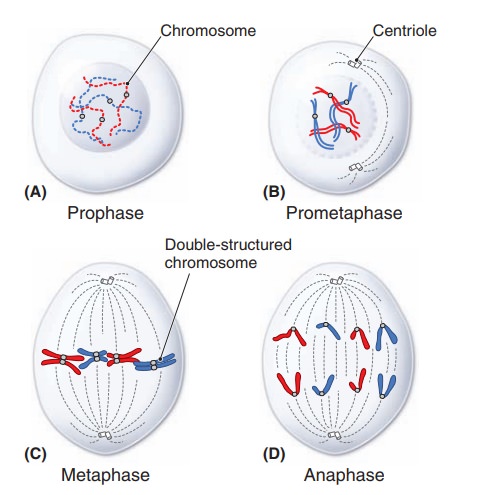
The goal of mitosis is to form
two daughter cells that have a complete set of genetic information. Mitosis is
divided into five stages: prophase, prometaphase, meta-phase, anaphase, and
telophase. During prophase the
chro-matin swells, or condenses, and the two sister chromatids are in close
approximation. The nucleolus disappears, and the mitotic spindle develops.
Spindle fibers start to form centrosomes, microtubule-organizing centers that
migrate to the poles of the cell. In prometaphase
the nuclear mem-brane vanishes and the chromosomes begin to disperse. They will
eventually attach to the microtubules that form the mitotic spindle. Metaphase is the stage of maximal
condensation. The chromosomes are in a linear formation in the center of the
cell, between the two spindle poles. It is during metaphase where cells can
most easily be analyzed to obtain a karyotype from an amniocentesis or
chorionic villus sampling (CVS). Anaphase
is initiated when the two chromatids separate. They form daughter chromosomes
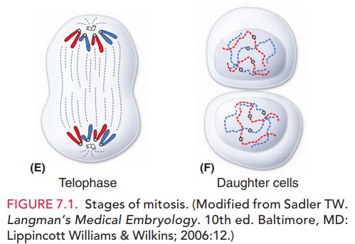
that are drawn to opposite poles of the cell by the spindle fibers. Finally, telophase is when the nuclear membrane
starts to reform around the independent daughter cells, which then go into
interphase (Fig. 7.1)
Meiosis
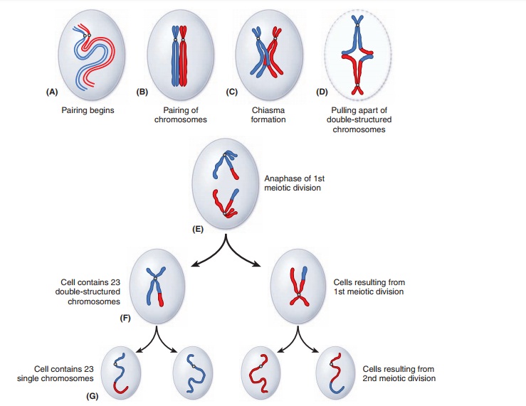
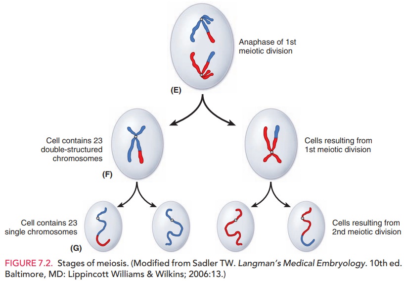
Meiosis differs
from mitosis in that a haploid number ofcells are initially produced in two
successive divisions. Thefirst division (meiosis I) istermed a
reduction division, because of the resulting decrease in chromosome number
from diploid to haploid. Meiosis I is also divided into four
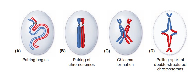
stages:prophase I, metaphase I, anaphase I, and telophase I. Prophase I is further divided into five
substages: lep-totene, zygotene, pachytene, diplotene, and diakinesis. In
prophase I the chromosomes condense and become shorter and thicker. It is
during the pachytene substage that crossing
over takes place, resulting in four distinct gametes. However, it is during
anaphase where most of the genetic variation occurs. In anaphase I the chromosomes go to opposite poles of the cell by independent assort-ment, signifying
that there are 223, or >8 million, possi-ble variations. Anaphase I is also the most error-prone
stepin meiosis. The process ofdisjunction,where
the chromo-somes go to opposite poles of the cell, can result in
non-disjunction, where both chromosomes go instead to the same pole. Nondisjunction is a common cause of
chromosomallyabnormal fetuses.
The second meiotic division (meiosis
II) is similar tomitosis, except that
the process occurs within a cell with a haploid number of chromosomes. Meiosis
II is also divided into fourstages: prophase II, metaphase II, anaphase II, and
telo-phase II. The result of meiosis II is four haploid daughter cells. After
anaphase II, the possibilities for genetic varia-tion are further increased by
223× 223,
ensuring genetic vari-ation (Fig. 7.2).
Related Topics