Chapter: Case Study in Obstetrics and Gynaecology: Emergency Gynaecology
Case Study Reports: Pain and the Intrauterine System
PAIN AND THE INTRAUTERINE SYSTEM
History
A
30-year-old woman had a levonorgestrel-releasing intrauterine system (IUS) inserted
by her general practitioner (GP) 3 weeks
ago. Ten days ago she presented to the emergency department with abdominal pain, and on examination the threads were not visible
and ultrasound scan suggested the IUS was misplaced in the right
uterine cornu. An appoint-
ment was made for hysteroscopic resection but
she
has presented again
in the interim
with further pain.
Examination
The
abdomen is not distended and is soft. There is generalized lower abdominal tender- ness. The threads cannot
be visualized on speculum examination.
Questions
·
How
would you explain
the symptoms and investigation findings?
·
How
would you further
investigate and manage this patient?
Answer:
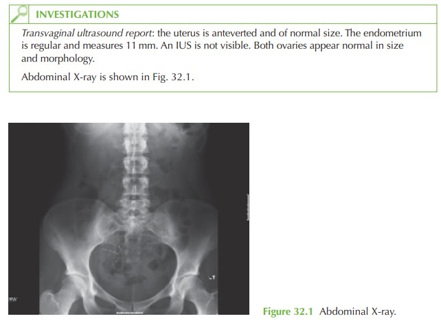
The
Plain X-ray shows
the IUS in the pelvis
but it is lying at a transverse angle in the right
pelvis. It is clearly not
within the uterus.
The current ultrasound result confirms that
the uterus is empty. However, the previous report suggested the device was at the uterine
cornu. It can be concluded therefore that the
device was inserted into the uterus
but it has subsequently migrated through the myometrium into the peritoneal cavity. We have no evidence to determine whether
or not it was originally placed in the
correct position at the
fundus.
Investigation
The
only important investigation is a pregnancy
test, as the woman is potentially preg- nant since the IUS
may not have
been effective if it was
never in the
correct location.
Management
The
IUS needs to be retrieved. While it was
in the uterus
this could have
been performed with outpatient hysteroscopic retrieval. However,
now a laparoscopy is indicated.
In
this case the laparoscopy revealed
blood-stained free fluid
in the pouch of Douglas, with scarring on the right fundal
area of the uterus. The IUS was found covered
with omentum in the
right lower abdomen. It was easily
removed laparoscopically.
As
the woman had wanted the IUS for contraception as well as treatment of her menor- rhagia, and as the
uterus appeared to have healed,
a new IUS was inserted under laparo-
scopic guidance at the time. Antibiotics were given to prevent infection.
Once an IUS or IUCD has
been inserted, women
should be advised
to have their
GP check the threads
are still visible
after the first
period. Thereafter, most women are willing and able to check the threads themselves.
Related Topics