Chapter: Medical Surgical Nursing: Assessment of Neurologic Function
Anatomy of the Central Nervous System
The Central Nervous System
ANATOMY OF THE BRAIN
The
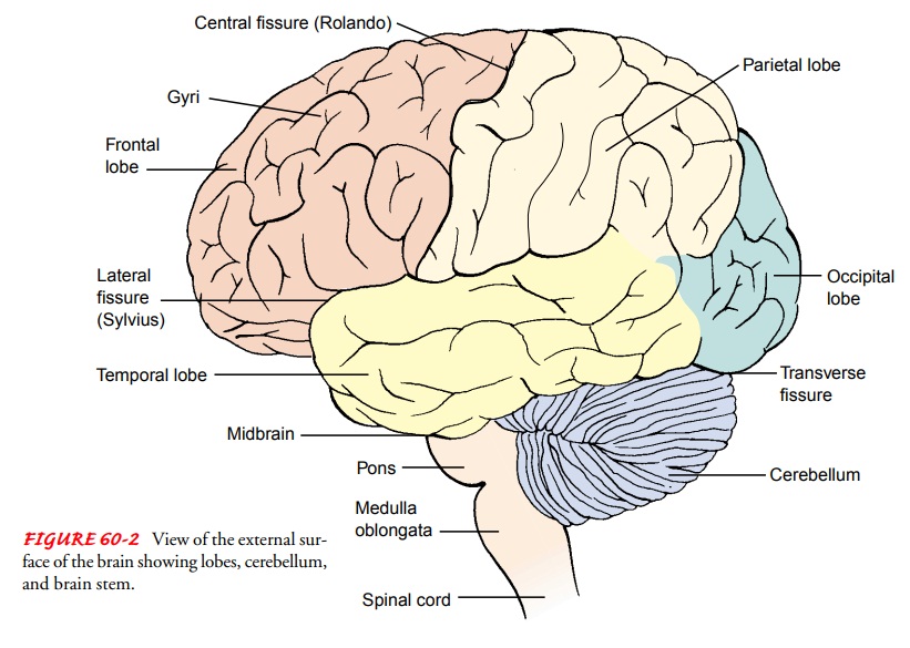
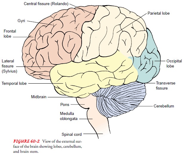
brain is divided into three major areas: the cerebrum, the brain stem, and the
cerebellum. The cerebrum is composed of two hemispheres, the thalamus, the
hypothalamus, and the basal gan-glia. Additionally, connections for the
olfactory (cranial nerve I) and optic (cranial nerve III) nerves are found in
the cerebrum. The brain stem includes the midbrain, pons, medulla, and
con-nections for cranial nerves II and IV through XII. The cerebel-lum is
located under the cerebrum and behind the brain stem (Fig. 60-2). The brain
accounts for approximately 2% of the total body weight; it weighs approximately
1,400 g in an average young adult (Hickey, 2003). In the elderly, the average
brain weighs approximately 1,200 g.
Cerebrum.
The cerebrum consists of two hemispheres that
areincompletely separated by the great longitudinal fissure. This sul-cus
separates the cerebrum into the right and left hemispheres. The two hemispheres
are joined at the lower portion of the fissure by the corpus callosum. The
outside surface of the hemi-spheres has a wrinkled appearance that is the
result of many folded layers or convolutions called gyri, which increase the
sur-face area of the brain, accounting for the high level of activity car-ried
out by such a small-appearing organ. The external or outer portion of the
cerebrum (the cerebral cortex) is made up of gray matter approximately 2 to 5
mm in depth; it contains billions of neurons/cell bodies, giving it a gray
appearance. White matter makes up the innermost layer and is composed of nerve
fibers and neuroglia (support tissue) that form tracts or pathways connect-ing
various parts of the brain with one another (transverse and as-sociation
pathways) and the cortex to lower portions of the brain and spinal cord
(projection fibers). The cerebral hemispheres are divided into pairs of
frontal, parietal, temporal, and occipital lobes. The four lobes are as follows
(see Fig. 60-2):
·
Frontal—the largest lobe. The
major functions of this lobe are concentration, abstract thought, information
storage or memory, and motor function. It also contains Broca’s area, critical
for motor control of speech. The frontal lobe is also responsible in large part
for an individual’s affect, judgment, personality, and inhibitions.
· Parietal—a predominantly sensory lobe. The primary sen-sory cortex, which analyzes sensory information and relays the interpretation of this information to the thalamus and other cortical areas, is located in the parietal lobe. It is also essential to an individual’s awareness of the body in space, as well as orientation in space and spatial relations.
·
Temporal—contains the auditory
receptive areas. Contains a vital area called the interpretive area that
provides inte-gration of somatization, visual, and auditory areas and plays the
most dominant role of any area of the cortex in cere-bration.
·
Occipital—the posterior lobe
of the cerebral hemisphere is responsible for visual interpretation.
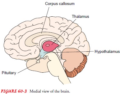
The corpus callosum (Fig. 60-3) is a thick collection of nerve fibers that connects the two hemispheres of the brain and is re-sponsible for the transmission of information from one side of the brain to the other. Information transferred includes sensation, memory, and learned discrimination. Right-handed people and some left-handed people have cerebral dominance on the left side of the brain for verbal, linguistic, arithmetical, calculating, and analytic functions. The nondominant hemisphere is responsible for geometric, spatial, visual, pattern, and musical functions.
The basal ganglia are masses of nuclei located deep
in the cere-bral hemispheres that are responsible for control of fine motor
movements, including those of the hands and lower extremities.
The thalamus (see Fig. 60-3) lies on either side of
the third ventricle and acts primarily as a relay station for all sensation
ex-cept smell. All memory, sensation, and pain impulses also pass through this
section of the brain.
The
hypothalamus is located anterior and inferior to the thal-amus. The
hypothalamus lies immediately beneath and lateral to the lower portion of the
wall of the third ventricle. It includes the optic chiasm (the point at which
the two optic tracts cross) and the mamillary bodies (involved in olfactory
reflexes and emotional response to odors). The infundibulum of the
hypo-thalamus connects it to the posterior pituitary gland. The hypo-thalamus
plays an important role in the endocrine system because it regulates the
pituitary secretion of hormones that influence metabolism, reproduction, stress
response, and urine production. It works with the pituitary to maintain fluid
balance and main-tains temperature regulation by promoting vasoconstriction or
vasodilatation.
The
hypothalamus is the site of the hunger center and is in-volved in appetite
control. It contains centers that regulate the sleep–wake cycle, blood
pressure, aggressive and sexual behavior, and emotional responses (ie,
blushing, rage, depression, panic, and fear). The hypothalamus also controls
and regulates the au-tonomic nervous system.
The
pituitary gland is located in the sella turcica at the base of the brain and is
connected to the hypothalamus. The pituitary is a common site for brain tumors
in adults; frequently they are de-tected by physical signs and symptoms that
can be traced to the pituitary, such as hormonal imbalance or visual
disturbances secondary to pressure on the optic chiasm.
Nerve
fibers from all portions of the cortex converge in each hemisphere and exit in
the form of a tight bundle of nerve fibers known as the internal capsule.
Having entered the pons and the medulla, each bundle crosses to the
corresponding bundle from the opposite side. Some of these axons make
connections with axons from the cerebellum, basal ganglia, thalamus, and hypo-thalamus;
some connect with the cranial nerve cells. Other fibers from the cortex and the
subcortical centers are channeled through the pons and the medulla into the
spinal cord.
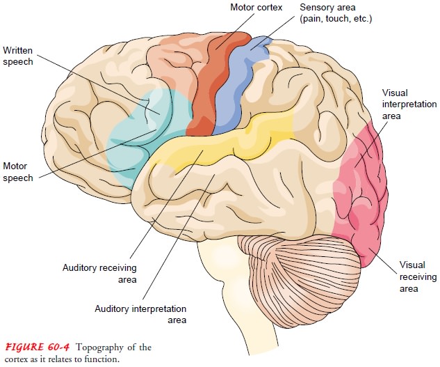
Although
the various cells in the cerebral cortex are quite sim-ilar in appearance,
their functions vary widely, depending on lo-cation. The topography of the
cortex in relation to certain of its functions is shown in Figure 60-4. The
posterior portion of each hemisphere (ie, the occipital lobe) is devoted to all
aspects of visual perception. The lateral region, or temporal lobe,
incorpo-rates the auditory center. The midcentral zone, or parietal zone,
posterior to the fissure of Rolando, is concerned with sensation; the anterior
portion is concerned with voluntary muscle move-ments. The large area behind
the forehead (ie, the frontal lobes) contains the association pathways that
determine emotional atti-tudes and responses and contribute to the formation of
thought processes. Damage to the frontal lobes as a result of trauma or dis-ease
is by no means incapacitating from the standpoint of mus-cular control or
coordination, but it affects a person’s personality, as reflected by basic
attitudes, sense of humor and propriety, self-restraint, and motivations.
Brain Stem.
The brain stem consists of the midbrain, pons, andmedulla oblongata (see
Fig. 60-2). The midbrain connects the pons and the cerebellum with
the cerebral hemispheres; it con-tains sensory and motor pathways and serves as
the center for au-ditory and visual reflexes. Cranial nerves III and IV
originate in the midbrain. The pons is situated in front of the cerebellum
be-tween the midbrain and the medulla and is a bridge between the two halves of
the cerebellum, and between the medulla and the cerebrum. Cranial nerves V through
VIII connect to the brain in the pons. The pons contains motor and sensory
pathways. Por-tions of the pons also control the heart, respiration, and blood
pressure.
The
medulla oblongata contains motor fibers from the brain to the spinal cord and
sensory fibers from the spinal cord to the brain. Most of these fibers cross,
or decussate, at this level. Cra-nial nerves IX through XII connect to the
brain in the medulla.
Cerebellum.
The cerebellum is separated from the cerebral hemi-spheres by a fold of
dura mater, the tentorium cerebelli. The cere-bellum has both excitatory and
inhibitory actions and is largely responsible for coordination of movement. It
also controls fine movement, balance, position
sense (awareness of where each part of the body is), and integration of
sensory input.
STRUCTURES PROTECTING THE BRAIN
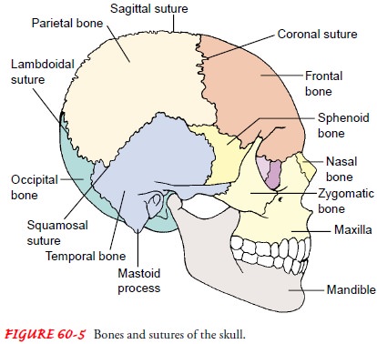
The
brain is contained in the rigid skull, which protects it from injury. The major
bones of the skull are the frontal, temporal, parietal, and occipital bones.
These bones join at the suture lines (Fig. 60-5).
The
meninges (fibrous connective tissues that cover the brain and spinal cord)
provide protection, support, and nourishment to the brain and spinal cord. The
layers of the meninges are the dura, arachnoid, and pia mater (Fig. 60-6).
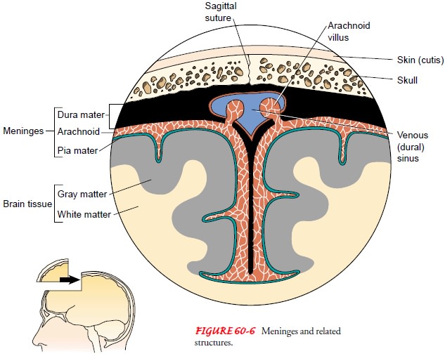
Dura mater—the outermost layer; covers the brain and the spinal cord. It is tough, thick, inelastic, fibrous, and gray.
There
are four extensions of the dura: the falx cerebri, which separates the two hemispheres
in a longitudinal plane; the tentorium, which is an infolding of the dura that
forms a tough membranous shelf; the falx cerebelli, which is be-tween the two
lateral lobes of the cerebellum; and the di-aphragm sellae, which provides a
“roof ” for the sella turcica. The tentorium supports the hemispheres and
separates them from the lower part of the brain. When excess pres-sure occurs
in the cranial cavity, brain tissue may be com-pressed against the tentorium or
displaced downward, a process called herniation. Between the dura mater and the
skull in the cranium, and between the periosteum and the dura in the vertebral
column, is the epidural space, a poten-tial space.
Arachnoid—the middle membrane; an extremely thin,
del-icate membrane that closely resembles a spider web (hence the name
arachnoid). It appears white because it has no blood supply. The arachnoid
layer contains the choroid plexus, which is responsible for the production of
cerebro-spinal fluid (CSF). This membrane also has unique finger-like
projections, arachnoid villi, that absorb CSF. In the normal adult,
approximately 500 mL of CSF is produced each day; all but 125 to 150 mL is
absorbed by the villi (Hickey, 2003). When blood enters the system (from trauma
or hemorrhagic stroke), the villi become obstructed and hy-drocephalus
(increased size of ventricles) may result. The subdural space is between the
dura and the arachnoid layer, and the subarachnoid space is located between the
arach-noid and pia layers and contains the CSF.
Pia
mater—the innermost membrane; a thin, transparent layer that hugs the brain
closely and extends into every fold of the brain’s surface.
CEREBROSPINAL FLUID
CSF, a clear and colorless fluid with a specific gravity of 1.007, is produced in the ventricles and is circulated around the brain and the spinal cord through the ventricular system. There are four ventricles: the right and left lateral, and the third and fourth ven-tricles. The two lateral ventricles open into the third ventricle at the interventricular foramen or the foramen of Monro. The third and fourth ventricles connect via the aqueduct of Sylvius. The fourth ventricle supplies CSF to the subarachnoid space and down the spinal cord on the dorsal surface. CSF is returned to the brain and is then circulated around the brain, where it is absorbed by the arachnoid villi.
CSF
is produced in the choroid plexus of the lateral, third, and fourth ventricles.
The ventricular and subarachnoid system con-tains approximately 125 to 150 mL
of fluid, while 15 to 25 mL of CSF is located in each lateral ventricle.
The
composition of CSF is similar to other extracellular flu-ids (such as blood
plasma), but the concentrations of the various constituents are different. The
analysis and laboratory report of CSF usually contains information on color,
specific gravity, pro-tein count, white blood cell count, glucose, and other
electrolyte levels; it may also be tested for immunoglobulins or lactate
(Hickey, 2003). Normal CSF contains a minimal number of white blood cells and
no red blood cells.
CEREBRAL CIRCULATION
The
cerebral circulation receives approximately 15% of the car-diac output, or 750
mL per minute. The brain does not store nu-trients and has a high metabolic
demand that requires the high blood flow. The brain’s blood pathway is unique
because it flows against gravity; its arteries fill from below and the veins
drain from above. In contrast to other organs that may tolerate de-creases in
blood flow because of their adequate collateral circula-tion, the brain lacks
additional collateral blood flow, which may result in irreversible tissue
damage when blood flow is occluded for even short periods of time.
Arteries.
Two internal carotid arteries and two vertebral arteriesand their extensive
system of branches provide the blood supply to the brain. The internal carotids
arise from the bifurcation of the common carotid and supply much of the
anterior circulation of the brain. The vertebral arteries branch from the
subclavian ar-teries, flow back and upward on either side of the cervical
verte-brae, and enter the cranium through the foramen magnum. The vertebral
arteries join to become the basilar artery at the level of the brain stem; the
basilar artery divides to form the two branches of the posterior cerebral
arteries. The vertebrobasilar arteries sup-ply most of the posterior
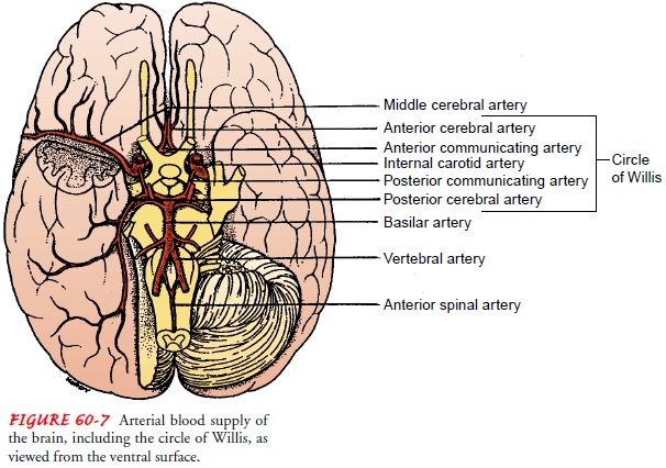
circulation of the brain.At the base of the brain
surrounding the pituitary gland, a ring of arteries is formed between the
vertebral and internal carotid ar-terial chains. This ring is called the circle
of Willis and is formed from the branches of the internal carotid arteries,
anterior and middle cerebral arteries, and anterior and posterior
communicat-ing arteries (Fig. 60-7). Functionally, the posterior portion of the
circulation and the anterior or carotid circulation usually remain separate.
The arteries of the circle of Willis can provide collateral circulation if one
or more of the four vessels supplying it become occluded or are ligated.
The arterial anastomoses along the circle of Willis
are frequent sites of aneurysms. These can be formed when the pressure at a
weakened arterial wall causes the artery to balloon out. Aneurysms may be
congenital or the result of degenerative changes in the vessel wall associated
with arteriosclerotic vascular disease. If an artery with an aneurysm bursts or
becomes occluded by vasospasm, an embolus, or a thrombus, the neurons distal to
the occlusion are deprived of their blood supply and the cells quickly die. The
result is a hemorrhagic stroke (cerebrovascular accident or in-farction). The
effects of the occlusion depend on which vessels are involved and which areas
of the brain these vessels supply.
Veins.
Venous drainage for the brain does not follow the
arterialcirculation as in other body structures. The veins reach the brain’s
surface, join larger veins, then cross the subarachnoid space and empty into
the dural sinuses, which are the vascular channels lying within the tough dura
mater (see Fig. 60-6). The network of the sinuses carries venous outflow from
the brain and empties into the internal jugular vein, returning the blood to
the heart. Cerebral veins and sinuses are unique because, unlike other veins in
the body, they do not have valves to prevent blood from flow-ing backward and
depend on both gravity and blood pressure.
BLOOD–BRAIN BARRIER
The CNS is inaccessible to many substances that circulate in the blood plasma (eg, dyes, medications, and antibiotics). After being injected into the blood, many substances cannot reach the neurons of the CNS because of the blood–brain barrier.
This barrier is formed by the
endothelial cells of the brain’s capillaries, which form continuous tight
junctions, creating a barrier to macromol-ecules and many compounds. All
substances entering the CSF must filter through the capillary endothelial cells
and astrocytes (Hickey, 2003). Often altered by trauma, cerebral edema, and
cerebral hypoxemia, the blood–brain barrier has implications in the treatment
and selection of medication for CNS disorders as well as serving a protective
function.
ANATOMY OF THE SPINAL CORD
The
spinal cord and medulla form a continuous structure ex-tending from the
cerebral hemispheres and serving as the con-nection between the brain and the
periphery. Approximately 45 cm (18 in) long and about the thickness of a
finger, it extends from the foramen magnum at the base of the skull to the
lower border of the first lumbar vertebra, where it tapers to a fibrous band
called the conus medullaris. Continuing below the second lumbar space are the
nerve roots that extend beyond the conus, which are called the cauda equina
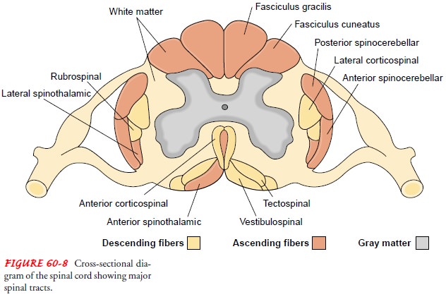
because they resemble a horse’s tail. Similar to the brain, the spinal cord
consists of gray and white matter. Gray matter in the brain is external and
white matter is internal; in the spinal cord, gray matter is in the center and
is sur-rounded on all sides by white matter (Fig. 60-8).
The spinal cord is surrounded by the meninges,
dura, arach-noid, and pia layers. Between the dura mater and the vertebral
canal is the epidural space. The spinal cord is an H-shaped struc-ture with
nerve cell bodies (gray matter) surrounded by ascend-ing and descending tracts
(white matter) (see Fig. 60-8). The lower portion of the H is broader than the
upper portion and cor-responds to the anterior horns. The anterior horns
contain cells with fibers that form the anterior (motor) root end and are
es-sential for the voluntary and reflex activity of the muscles they
in-nervate. The thinner posterior (upper horns) portion contains cells with
fibers that enter over the posterior (sensory) root end and thus serve as a
relay station in the sensory/reflex pathway.
The thoracic region of the spinal cord has a
projection from each side at the crossbar of the H of gray matter called the
lateral horn. It contains the cells that give rise to the autonomic fibers of
the sympathetic division. The fibers leave the spinal cord through the anterior
roots in the thoracic and upper lumbar segments.
Sensory and Motor Pathways: The Spinal Tracts.
The whitematter of the
cord is composed of myelinated and unmyelinated nerve fibers. The
fast-conducting myelinated fibers form bun-dles that also contain glial cells.
Fiber bundles with a common function are called tracts. There are six ascending
tracts. Two conduct sensation, principally the perception of touch, pres-sure,
vibration, position, and passive motion from the same side of the body. Before
reaching the cerebral cortex, these fibers cross to the opposite side in the
medulla. The two spinocere-bellar tracts conduct sensory impulses from muscle
spindles, providing necessary input for coordinated muscle contraction. They
ascend essentially uncrossed and terminate in the cerebel-lum. The last two
spinothalamic tracts are responsible for con-duction of pain, temperature,
proprioception, fine touch, and vibratory sense from the upper body to the
brain. They ascend, cross to the opposite side of the brain, and terminate in
the thal-amus (Hickey, 2003).
There
are eight descending tracts, seven of which are engaged in motor function. The
two corticospinal tracts conduct motor impulses to the anterior horn cells from
the opposite side of the brain and control voluntary muscle activity. The three
vestibu-lospinal tracts descend uncrossed and are involved in some autonomic
functions (sweating, pupil dilation, and circulation) and involuntary muscle
control. The corticobulbar tract conducts impulses responsible for voluntary
head and facial muscle move-ment and crosses at the level of the brain stem.
The rubrospinal and reticulospinal tracts conduct impulses involved with
invol-untary muscle movement.
Vertebral Column.
The bones of the vertebral column surroundand protect the spinal cord and normally consist of 7 cervical, 12 thoracic, and 5 lumber vertebrae, as well as the sacrum (a fused mass of five vertebrae), and terminate in the coccyx. Nerve roots exit from the vertebral column through the intervertebral foramina (openings). The vertebrae are separated by disks, ex-cept for the first and second cervical, the sacral, and the coccygeal vertebrae. Each vertebra has a ventral solid body and a dorsal seg-ment or arch, which is posterior to the body. The arch is com-posed of two pedicles and two laminae supporting seven processes. The vertebral body, arch, pedicles, and laminae all en-case the vertebral canal.
Related Topics