Chapter: 11th 12th standard bio zoology Human Body higher secondary school
Anatomy of Nephrolepsis

Nephrolepsis
Division : Tracheophyta
Sub division : Pteropsida
Class : Leptosporangiatae
Order : Filicales
Family : Dennstaedtiaceae
Genus : Nephrolepsis
The genus Nephrolepsis is a tropical fern with about 30 species. Most of the species are found in terrestrial habitats. Some species are epiphytes e.g., N. volubilis and N. ramosa. Several species are grown as ornamental plants. In India, there are 5 species. Of these,N. acuta and N. tuberosa are common species.
Anatomy
Rhizome :
The Rhizome is differentiated into epidermis, hypodermis, ground tissue and stele. The stele is a meristele (Fig) A meristele is a part of dictyostele found between two neighbouring leaf gaps and appear as separate strand in a transverse section. A dictyostele is a solenostele with leaf gaps and distinct vascular strands. A solenostele is a condition when a mass of parenchyma cells found in the centre of the xylem. The epidermis is the protective layer with a thick layer of cuticle. The hypodermis is more or less continuous and heavily sclerotic. This region is followed by parenchymatous ground tissue with starch grains.
The stele structure varies within the same rhizome. A mature rhizome with many leaves has dictyostele which gets separated into a number of strands called meristeles. Each meristele is surrounded by its own endodermis which is followed by pericycle. The pericycle is followed by phloem. The central region of stele is occupied by xylem.
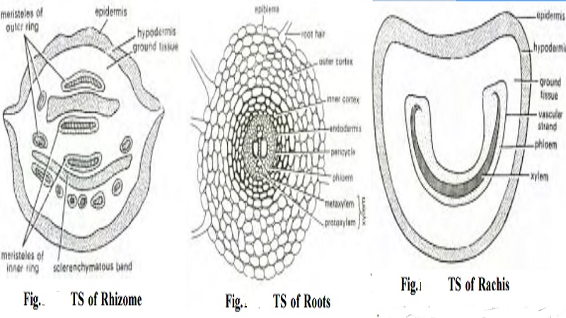
Root: The transverse section of root has three distinct parts - epiblema, cortex and vascular cylinder (fig). The epiblema is the outermost layer of thin walled cells.
Some cells of this region produce unicellular root hairs. The cortex is divided into outer parenchymatous and inner sclerenchymatous regions. The latter provides machanical support to roots. The innermost region of cortex has endodermis. Next to this layer is pericycle.
The vascular cylinder is diarch and exarch. A diarch condition consists of two protoxylem points. An exarch condition refers to presence of protoxylem away from the centre of the axis.
Rachis or Petiole: The transverse section of rachis has epidermis, hypodermis, ground tissue and stele (fig). The epidermis consists of a single layer of cells with cuticle. This layer is followed by 2- 3 layered sclerenchymatous hypodermis. Next to this is a broad zone of parenchymatous ground tissue. At the centre, a 'U' shaped meristele is located. This stele is similar to that of rhizome.
Pinna: The internal structure resembles a dicot leaf. The upper and lower epidermis are single layered. The outer walls of both epidermis have thick cuticle. The mesophyll region is differentiated into a columnar palisade parenchyma and loosely arranged spongy parenchyma cells. A concentric vascular bundle is found in the centre with a distinct bundle sheath.
Related Topics