Chapter: Basic Radiology : Liver, Biliary Tract, and Pancreas
Techniques and Normal Anatomy - Liver, Biliary Tract, and Pancreas
TECHNIQUES AND
NORMAL ANATOMY
Several modalities such as US,
nuclear medicine, CT, and MR imaging are commonly used in diagnosing diseases
of liver, pancreatic, or biliary ductal system.
With US, normal organs are
displayed as structures of different echogenicity. In general, fluid is
anechoic (has noechoes). Soft tissue has echoes of mild to moderate intensity.
Bone has extremely strong echoes. Abnormal organs are dis-played as areas of
diffuse inhomogeneity or as focal regions of decreased or increased
echogenicity within the organ. The normal appearances of the liver, biliary
system, and pancreas have been well established. Echogenicity of the organs in
the abdomen is evaluated in relation to other nearby organs. The pancreas is typically
the most echogenic organ in the upper abdomen, followed by the liver. The liver
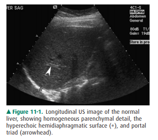
typically has homo-geneous parenchymal detail (Figure 11-1). Numerous
intra-hepatic vessels including portal veins and hepatic veins are easily seen
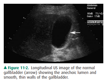
within the liver. The gallbladder appears as an anechoic pear-shaped structure
along the inferior aspect of the liver (Figure 11-2). It normally has a thin,
homogeneous wall less than 3 mm in thickness. The degree of distention ofthe
gallbladder varies with postprandial intervals. As is ex-pected, it contracts
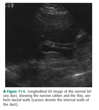
after a meal and distends in the fasting state. The biliary ducts are thin
tubes, the walls of which are 1.5 mm or less (essentially unmeasurable). The
ducts in-crease in caliber as they extend from the liver to the sphincter of
Oddi (Figure 11-3). The upper limit in caliber of the ex-trahepatic biliary
ducts increases with age. When measured at the level where it crosses the right
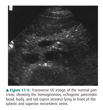
hepatic artery, 6 mm is usually used as the cutoff diameter. The pancreas is
homoge-neous, comma-shaped, and parallel to the splenic vein and extends from
the left upper quadrant caudally and to the right (Figure 11-4). In
anteroposterior dimension the pan-creatic head is approximately 3 cm, the body
2.5 cm, and the tail 2 cm. The pancreas can sometimes be difficult to image
with ultrasound because of its relatively posterior position and overlying
bowel gas. The normal pancreatic duct, if seen, should be 3 mm or less.
With NM, normal organs are
displayed as regions of ho-mogeneous activity conforming to the general shape
of the organ. Abnormal organs are displayed as diffuse inhomo-geneity or as
focal areas of reduced or increased activity. In the past, the liver was most
commonly studied with NM with technetium-labeled sulfur colloid. However, this
technique has largely been replaced by CT, US, and MR imaging. The most common
NM study of the liver today utilizes tech-netium-labeled red blood cells to
evaluate for cavernous he-mangioma. Evaluation of the biliary system is a
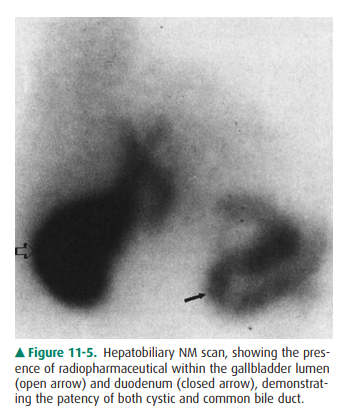
common application for NM studies. Technetium-labeled hepatobiliaryimaging
iminodiacetic acid derivatives for hepatobiliary im-aging, especially disofenin
and mebrofenin, are taken up by the liver, excreted into the bile, carried to
the biliary tree and gallbladder, and from there travel to the bowel through
the extrahepatic ducts (Figure 11-5). Depending on the exact agent used, these
are termed hepatic iminodiacetic acid, HIDA scans. Currently, no practical
imaging of the pancreas is done by means of NM techniques.
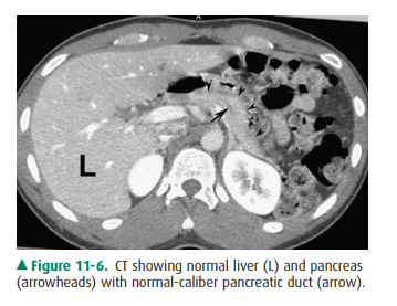
With CT, normal organs are
displayed as regions of differ-ing attenuation. Abnormal organs are displayed
as diffuse in-homogeneity or as focal areas of decreased or increased
attenuation. The liver, biliary system, and pancreas are well demonstrated by
CT (Figure 11-6). Intravenous contrast aids in their evaluation. The liver is
the most dense organ in the abdomen. The normal liver parenchyma appears
homoge-neous, just as in US. The portal and hepatic vessels and the biliary
ductal system are likewise easy to identify. Overall measurements of wall
thickness and biliary duct caliber are the same as for US. The pancreas is
easily identified on CT, and the pancreatic duct is frequently well seen.
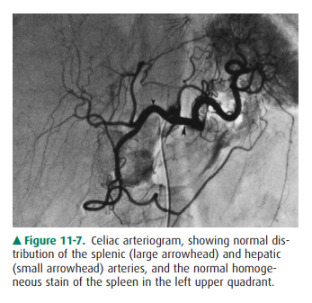
At angiography, normal organs
enhance to variable ex-tents. Abnormal organs either inhomogeneously enhance or
have focal areas of decreased or increased enhancement. Although the parenchyma
of the normal organs is rarely demonstrated, the blood vessels of these organs
are seen in exquisite detail (Figure 11-7). In the liver, both the hepatic
artery and all of its branches can be seen. Delayed studies through the liver
in the venous phase demonstrate the por-tal vein. The cystic artery and any
collateral vessels can beangiographically demonstrated. Angiographic studies of
the pancreas can demonstrate major pancreatic branches, as well as encasement,
displacement, stenosis, or occlusion.
On MR imaging, normal organs have
homogeneous sig-nal intensity or well-recognized variations in signal
inten-sity. Abnormal organs have inhomogeneous signal intensity or areas of
increased or decreased signal intensity. The normalliver, biliary system, and
pancreas are well demonstrated on MR imaging (Figure 11-8). The liver has a
homogeneous signal intensity which is usually higher than that of muscle and
lower than that of the spleen. The biliary system is nor-mally demonstrated as
an area of low signal intensity on T1-weighted images and high signal intensity
on T2-weighted images. This appearance reflects the fluid bile within the
gallbladder and biliary tree. Magnetic resonance cholangiopancreatography, or
MRCP, demonstrates the bil-iary system as very high signal intensity structures
against a very low signal intensity background of surrounding solid tissues
(Figure 11-9). The pancreas is of intermediate signal on both T1- and
T2-weighted images and may be hard to differentiate from bowel if no oral
contrast agent is admin-istered to the patient. As in CT and US, the normal
fatty change within the pancreas that occurs with age is visible. Newer hepatocyte-specific
contrast agents, such as gadoxe-tate disodium, are offering new ways of
evaluating the liver and biliary system.


Related Topics