Chapter: 11th Botany : Chapter 6 : Cell: The Unit of Life
Microscopy and its types
Microscopy
Microscope is an inevitable
instrument in studying the cell and subcellular structures. It offers scope in
studying microscopic organisms therefore it is named as microscope (mikros –
small; skipein – to see) in Greek terminology. Compound microscope was invented
by Z. Jansen.
Resolution: The term resolving power or resolution refers to the
ability of the lenses to show the details of object lying between two points.
It is the finest detail available from an object. It can be calculated using
the following formula
Resolution = 0.61λ/NA
Where, λ= wavelength of the light and
NA is the numerical aperture.
Numerical Aperture: It is an important optical constant associated with the
optical lens denoting the ability to resolve. Higher the NA value greater will
be the resolving power of the microscope.
Magnification: The optical
increase in the size of an image is
called magnification. It is calculated by the following formula
Magnification = size of image seen
with the microscope / size of the image seen with normal eye
Microscope works on the lens
system which basically relies on properties of light and lens such as
reflection, magnification and numerical aperture. The common light microscope
which has many lenses are called as compound
microscope. The microscope transmits visible light from sources to eye or
camera through sample, where interaction takes place.
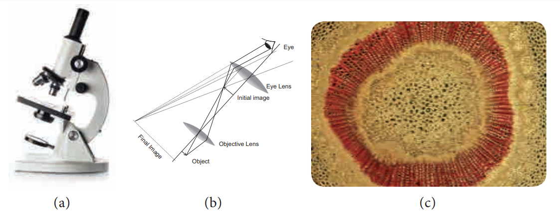
1. Bright field Microscope
Bright field microscope is routinely used
microscope in studying various aspects of cells. It allows light to pass
directly through specimen and shows a well distinguished image from different
portions of the specimen depending upon the contrast from absorption of visible
light. The contrast can be increased by staining the specimen with reagent that
reacts with cells and tissue components of the object.
The light rays are focused by condenser on to the
specimen on a microslide placed upon the adjustable platform called as stage. The light comes from the Compact
Flourescent Lamp (CFL) or Light Emitting Diode (LED) light system. Then it
passes through two lens systems namely objective lens (closer to the object)
and the eye piece (closer to eye). There are four objective lenses (5X, 10X,
45X and 100X) which can be rotated and fixed at certain point to get required
magnification. It works on the principle of numerical aperture value and its
own resolving power.
The first magnification of the microscope is done
by the objective lens which is called primary
magnification and it is real, inverted image. The second magnification of
the microscope is obtained through eye piece lens called as secondary magnification and it is
virtual and inverted image (Figure 6.2 a, b and c).
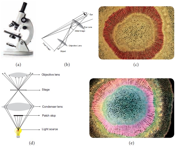
2. Dark Field Microscope
The dark field microscope was discovered by Z. Sigmondy (1905). Here the field will
be dark but object will be glistening so the appearance will be bright. A
special effect in an ordinary microscope is brought about by means of a special
component called ‘Patch Stop Carrier’.
It is fixed in metal ring of the condenser component. Patch stop is a small
glass device which has a dark patch at centre of the disc leaving a small area
along the margin through which the light passes. The light passing through the
margin will travel oblique like a hollow cone and strikes the object in the
periphery, therefore the specimen appears glistening in a dark background.
(Figure 6.2 d and e).
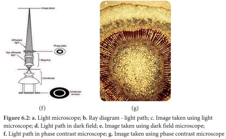
3. Phase contrast microscope
This was invented by Zernike (1935). It is a modification of
light microscope with all its basic principle. The objects observed by
increasing the contrast by bringing about change in amplitude of the light
waves. The contrast can be increased by introducing the ‘Phase Plate’ in the condenser lens. Phase plate is a circular
component with circular annular etching.
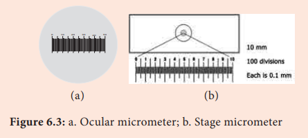
Microscopic measurements:
The microscope also has facility to
measure microscopic objects through a technique called ‘micrometry’. There are two scales involved for measuring.
1.
Ocular Micrometer
2.
Stage Micrometer
Ocular Micrometer: It is fixed inside
the eye piece lens. It is a thin transparent glass disc where there are lines divided into 100 equal units. The scale
has no value.
Stage Micrometer: This is a slide
with a line divided into 100 units. The line is about 1mm. The distance between two adjacent lines is 10 µm. The known
value of the stage micrometer is transferred to the ocular micrometer, thereby
the measurements can be made using ocular micrometer.
Light passes with different
velocity after coming out of the thinnest and thickest areas of the phase plate
thereby increasing the contrast of the specimen. A hollow cone of light passes
through the condenser. Direct light pass through thin area of phase plate,
whereas light passing from the specimen reaches thick area of phase plate. The
light passing through thicker area travel at low speed, on the other hand the
light passing through thin area reach fast therefore contrast is increased in
the specimen. Phase contrast microscope is used to observe living cells,
tissues and the cells cultured invitro during
mitosis (Figure 6.2 f and g).
4. Electron Microscope
Electron Microscope was first introduced by Ernest Ruska (1931) and developed by G Binning and H Roher (1981). It is used to analyse the fine details of the cell
and organelles called ultrastructure. It uses beam of accelerated electrons as
source of illumination and therefore the resolving power is 1,00,000 times than
that of light microscope.
The specimen to be viewed under electron microscope
is dehydrated and impregnated with electron opaque chemicals like gold or
palladium. This is essential for withstanding electrons and also for contrast of the image.
There are two kinds of electron microscopes namely
1.
Transmission Electron Microscope (TEM)
2.
Scanning Electron Microscope (SEM)
1. Transmission electron microscope:
Transmission
electron microscope: This is the most commonly used electron microscope
which provides two dimensional image. The components of the microscope are as
follows:
![]()
![]()
![]()
a.
Electron Generating System
b.
Electron Condensor
c.
Specimen Objective
d.
Tube Lens
e.
Projector
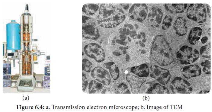
A beam of electron passes through the specimen to
form an image on fluorescent screen. The magnification is 1–3 lakhs times and
resolving power is 2–10 Å. It is used for studying detailed structrue of
viruses, mycoplasma, cellular organelles, etc (Figure 6.4 a and b).
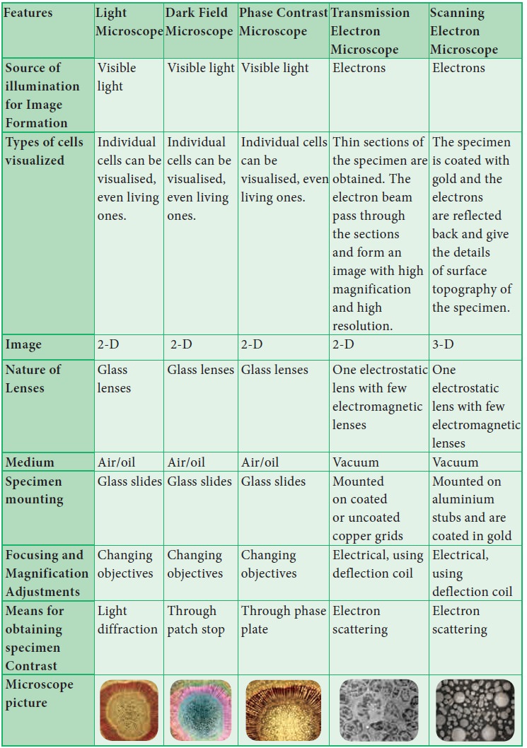
Comparison of Microscopes
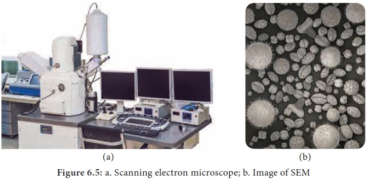
2. Scanning Electron Microscope:
This is used to obtain three dimensional image and
has a lower resolving power than TEM. In this, electrons are focused by means
of lenses into a very fine point. The interaction of electrons with the
specimen results in the release of different forms of radiation (such as auger
electrons, secondary electrons, back scattered electrons) from the surface of
the specimen. These radiations are then captured by an appropriate detector,
amplified and then imaged on fluorescent screen. The magnification is 2,00,000
times and resolution is 5–20 nm (Figure 6.5 a and b).
Related Topics