Chapter: Microbiology and Immunology: Virology, Virus: Herpesviruses
Laboratory Diagnosis - Herpes Simplex Virus Infections
Laboratory Diagnosis
◗ Specimens
These include saliva, vesicle fluid, conjunctival fluid, corneal scraping, skin swab, skin scrapings, and cerebrospinal fluid (CSF).
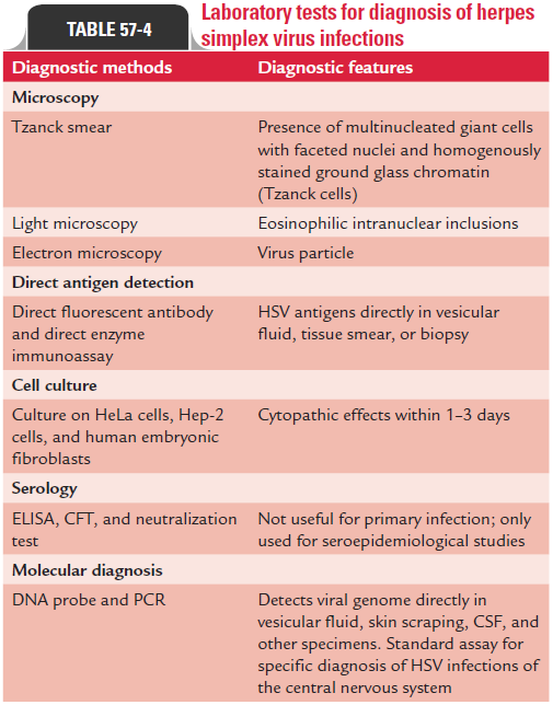
◗ Microscopy
Light microscopy of the stained infected cell may show bal-looning of cells, ground glass nuclei and eosinophilic intra-nuclear inclusions, and multinucleated giant cells. Electron microscopy can be used for direct demonstration of virions in the negatively stained smears of the clinical specimens.
◗ Direct antigen detection
Direct enzyme immunoassay and direct fluorescent anti-body test are useful to demonstrate HSV antigens directly in vesicular fluid, tissue smear, or biopsy.
◗ Isolation of the virus
A definitive diagnosis of HSV infection is made by isolating the viruses in cell cultures.
Cell culture: For culture, scrapings of skin vesicles and muco-sal lesions are collected, transferred immediately in a vial trans-port medium, and are carried to microbiology laboratory. After inoculation, HSV produces cytopathic effects (CPEs) within 1–3 days on HeLa cells, Hep-2 cells, and human embryonic fibroblasts. These cells become enlarged and appear ballooned. Some virus strains, particularly HSV-2, cause fusion of infected cells, leading to formation of syncytium. Immunofluorescent staining of infected tissue culture is useful to confirm the diag-nosis within 24 hours. Polymerase chain reaction (PCR) is a use-ful tool to distinguish HSV-1 from HSV-2. Isolated HSV can be typed by biochemical, immunological, and molecular methods.
◗ Serodiagnosis
Serodiagnosis is of little value for diagnosis of primary HSV infection. It is mainly used for epidemiological studies. It is used only to determine postexposure to HSV. It is not useful for diagnosing recurrent infections because rise in antibody titer does not usually correlate with recurrent disease. Also, the serological tests cannot distinguish between HSV-1 and HSV-2 antibodies due to cross-reactivity.
◗ Other tests
Histology
Tzanck smear: It is useful for the cytological identificationof viruses in the scraping obtained from the base of the vesicular lesion. In this procedure, the vesicular lesion is aseptically ruptured, and the base of the lesion is scrapped with a scalpel. The scraping from the base of the lesion is smeared on a glass slide, air dried, fixed, and usually stained with Giemsa or Wright stain. Demonstration of typical giant cell or Cowdry type A intranuclear inclusion bodies in the stained smear is diagnostic of HSV infection. This method shows variable sensitivity of 40–80% for diagnosis of HSV. Laboratory tests for diagnosis of HSV infections are summarized in Table 57-4.
Related Topics