Chapter: Basic Radiology : The Physical Basis of Diagnostic Imaging
Interaction of X-rays with Matter
Interaction of
X-rays with Matter
X-rays primarily interact with
matter through interaction of their oscillating electric field with the atomic
electrons in the material. Having no electrical charge, the x-rays are more
penetrating than other types of ionizing radiation (such as alpha or beta
particles) and are therefore useful for imaging the human body. The x-rays may
be absorbed or scattered by the atomic electrons. In the absorption process
(photoelectric absorption), the x-ray is completely absorbed, giving all its
energy to an inner shell atomic electron, which is then ejected from the atom
and goes on to ionize other atoms in the im-mediate vicinity of the initial
interaction. In the scattering process (Compton scattering), the x-ray
ricochets off an atomic electron, losing some of its energy and changing its
direction. The recoiling electron also goes on to ionize hun-dreds of atoms in
the vicinity. Electrons from both processesgo on to ionize many other atoms and
are responsible for the biological damage produced by x-rays.
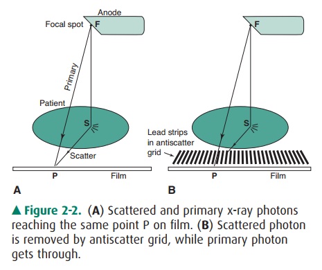
Figure 2-2. (A) Scattered and primary x-ray photons reaching the same point P on film. (B) Scattered photon is removed by antiscatter grid, while primary photon gets through.
The attenuation of the x-ray
intensity with thickness of material follows an exponential law due to the
random hit-or-miss nature of the interaction. The process is similar to fir-ing
a volley of rifle bullets into a forest, where the bullets may either stick in
a tree (be absorbed) or ricochet off a tree (scat-ter). The deeper you go into
the forest, the fewer bullets there are; however, a bullet still has a chance
of traveling through the forest without hitting a tree. Likewise, an x-ray can
make it all the way through a patient’s body without touching any-thing and
remain unchanged, as if it had passed through a vacuum instead. These are
called primary x-rays. Typically, only about 1% of the incident x-rays
penetrate the patient, and only about a third of these are primary x-rays; the
rest are scattered x-rays that do not contribute to the anatomic image. An
x-ray image is a shadow or projection image which assumes that x-rays reaching
the film have traveled in a straight line from the source, but this is true
only for the pri-mary x-rays. As Figure 2-2A shows, the film density
(black-ness) at point P on the film is related to the anatomy along line FP.
The scattered photon reaches the film along the path FSP and is relaying
information about the anatomy at the random point S to point P on the film.
Scatter simply pro-duces a uniform gray background; it does not contribute to
the image. Because scatter reduces image contrast, it is desir-able that the
scatter be removed. This task is accomplished by use of an antiscatter grid
(Figure 2-2B). This grid consists of a series of narrow lead strips with
radiolucent (low-attenuation) interspace material to remove some of the
scatter. With the grid, the scattered photon shown in the figure can no longer
reach the film, but the primary x-rays can. More of the scat-ter than primary
x-rays is eliminated by the grid; hence, image contrast increases, but at the
cost of an increase of a factor of 2 to 3 in patient dose. This increase occurs
because the scatter, which was previously blackening the film, has been
reduced, and therefore, higher x-ray exposure to the front of the patient is
necessary to get the requisite number of x-rays through the grid to blacken the
film. The grid is usu-ally made to move a few interspaces during the exposure
by a motor drive, in order to wash out the grid lines.
The absorption process is more
prevalent at lower kilo-voltages and in materials with higher atomic numbers.
Bones appear white on an x-ray film because photoelectric absorp-tion of x-rays
is greater in bone than in soft tissue as a result of the higher atomic number
of bone. Lead is a useful shield-ing material for x-rays because of its high
atomic number. The probability of the absorption process decreases rapidly with
photon energy (as 1/E3) and the scattering process de-creases slowly
(as 1/E); hence, the x-ray beam becomes more penetrating as kilovoltage
increases. The scattering process is roughly independent of the atomic number
of the attenuat-ing material (all electrons look alike to the photon for the
scattering process), whereas the absorption process is more probable for
tightly bound electrons such as the inner elec-trons in heavier elements.
Increasing the kilovoltage is
therefore beneficial to the pa-tient in that it reduces the radiation dose:
that is, fewer x-rays must penetrate the front of the patient to get the
requisite number out the back to blacken the film. However, an in-crease in
kilovoltage will reduce image contrast because the absorption process, which is
sensitive to atomic number, will decrease and the scattering process is
independent of the atomic number of the materials. Even with materials of the
same atomic number, contrast improves at lower kilovoltage settings because of
higher attenuation, which results in greater differential attenuation between
different thicknesses of the same material. Thus there is a tradeoff between
image quality (contrast) and patient dose that must be weighed in the selection
of kilovoltage.
Related Topics