Zoology - Human excretory system | 11th Zoology : Chapter 8 : Excretion
Chapter: 11th Zoology : Chapter 8 : Excretion
Human excretory system
Human excretory system
1. Structure of kidney
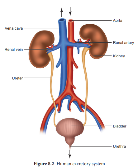
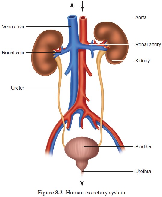
Excretory system in human consists of a pair of
kidneys, a pair of ureters, urinary bladder and urethra (Figure. 8.2). Kidneys
are reddish brown, bean shaped structures that lie in the superior lumbar
region between the levels of the last thoracic and third lumber vertebra close
to the dorsal inner wall of the abdominal cavity. The right kidney is placed
slightly lower than the left kidney. Each kidney weighs an average of 120-170
grams. The outer layer of the kidney is covered by three layers ofsupportive
tissues namely, renal fascia, perirenal fat capsule and fibrous capsule.
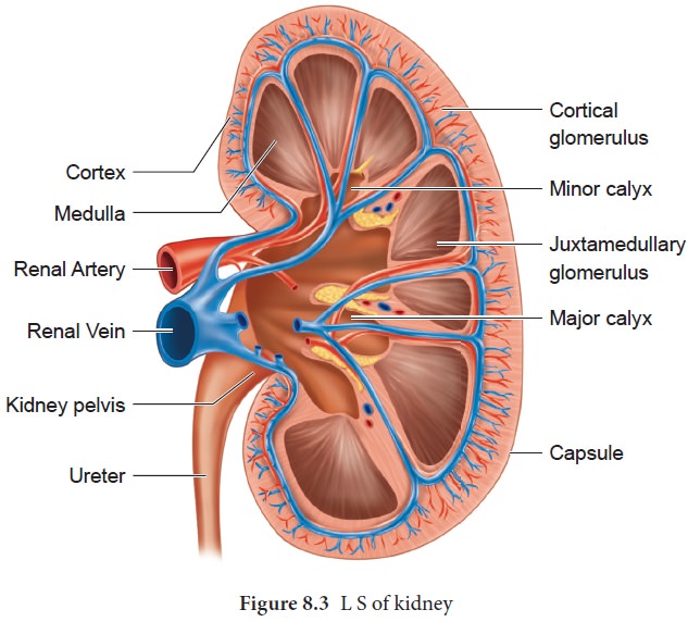
The longitudinal section of kidney (Figure. 8.3)
shows, an outer cortex, inner medulla and pelvis. The medulla is divided into a
few conical tissue masses called medullary pyramids or renal pyramids. The part
of cortex that extends in between the medullary pyramids is the renal columns
of Bertini. The centre of the inner concave surface of the kidney has a notch
called the renal hilum, through which ureter, blood vessels and nerves
innervate. Inner to the hilum is a broad funnel shaped space called the renal pelvis
with projection called calyces.
The renal pelvis is continuous with the ureter once it
leaves the hilum. The walls of the calyces, pelvis and ureter have smooth
muscles which contracts rhythmically. The calyces collect the urine and empties
into the ureter, which is stored in the urinary bladder temporarily. The
urinary bladder opens into the urethra through which urine is expelled out.
2. Structure of a nephron
Each kidney has nearly one million complex tubular
structures called nephron (Figure 8.4). Each nephron consists of a filtering
corpuscle called renal corpuscle (malpighian body) and a renal tubule.
The renal tubule opens into a longer tubule called the collecting duct. The
renal tubule beginswith a double walled cup shaped structure called the
Bowman’s capsule, which encloses a ball of capillaries that delivers fluid to
the tubules, called the glomerulus (Figure 8.4). The Bowman’s capsule and the
glomerulus together constitute the renal
corpuscle. The endothelium of glomerulus has many pores (fenestrae). The
external parietal layer of the Bowman's capsule is made up of simple squamous
epithelium and the visceral layer is made of epithelial cells called podocytes.
The podocytes end in foot processes which cling to the basement membrane of the
glomerulus. The openings between the foot processes are called filtration
slits.
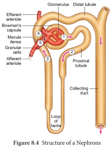
The renal tubule continues further to form the proximal convoluted tubule [PCT] followed by a U-shaped loop of Henle (Henle’s
loop) that has a thin descending and a thick ascending limb. The ascending limb
continues as a highly coiled tubular region called the distal convoluted tubule
[DCT]. The DCT of many nephrons open into a straight tube called collecting
duct. The collecting duct runs through the medullary pyramids in the region of
the pelvis. Several collecting ducts fuse to form papillary duct that delivers
urine into the calyces, which opens into the renal pelvis.
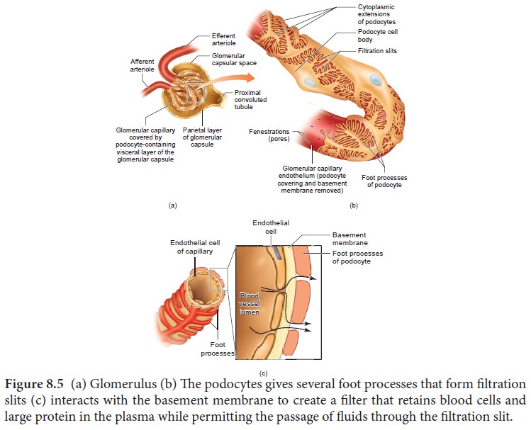
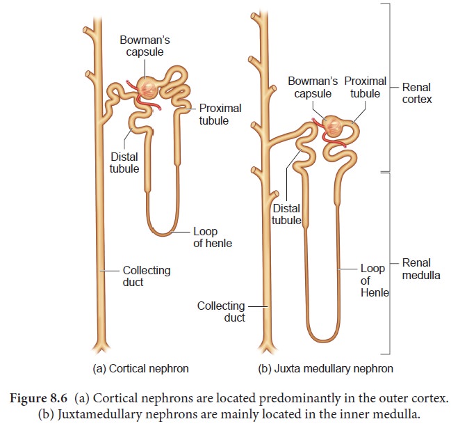
In the renal tubules, PCT and DCT of the nephron are situated in the cortical region of the kidney whereas the loop of Henle is in the medullary region. In majority of nephrons, the loop of Henle is too short and extends gives several foot processes that form filtration slits (c) interacts with the basement membrane to create a filter that retains blood cells and large protein in the plasma while permitting the passage of fluids through the filtration slit.
only very little into the medulla and are called
cortical nephrons. Some nephrons have very long loop of Henle that run deep
into the medulla and are called juxta
medullary nephrons (JMN) (Figure
8.6 a and b)
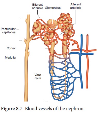
The
capillary bed of the nephrons-First capillary bed of the
nephron is the glomerulus and the
other is the peritubular capillaries. The glomerular capillary bed is different
from other capillary beds in that it is supplied by the afferent and drained by
the efferent arteriole. The efferent arteriole that comes out of the glomerulus
forms a fine capillary network around the renal tubule called the peritubular
capillaries. The efferent arteriole serving the juxta medullary nephron forms bundles of long straight vessel called vasa recta
and runs parallel to the loop of Henle.
Vasa recta is absent or reduced in cortical
nephrons (Figure 8.7).
Related Topics