Chapter: Ophthalmology: Conjunctiva
Conjunctiva: Basic Knowledge
Conjunctiva
Basic Knowledge
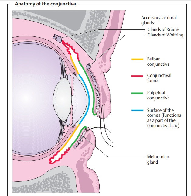
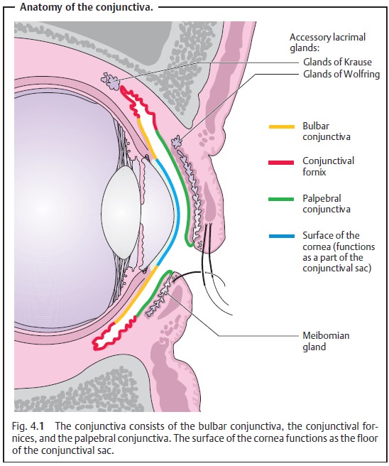
Structure of the conjunctiva (Fig. 4.1): The
conjunctiva is a thin vascularmucous membrane that normally of shiny
appearance. It forms the conjunc-tival sac together with the surface of the
cornea. The bulbar conjunctiva is
loosely attached to the sclera and is more closely attached to the limbus of
the cornea. There the conjunctival epithelium fuses with the corneal
epithelium. The palpebral conjunctiva
lines the inner surface of the eyelid and is firmly attached to the tarsus. The
loose palpebral conjunctiva forms a fold in the conjunctival fornix, where it joins the bulbar conjunctiva. A
half-moon-shaped fold of mucous membrane, the plica semilunaris, is located in
the medial corner of the palpebral fissure. This borders on the lacrimal
caruncle, which contains hairs and sebaceous glands.
Function of the conjunctival sac: The conjunctival sac has three main tasks:
1. Motility of the
eyeball.The loose
connection between the bulbar conjunc-tiva and the sclera and the “spare”
conjunctival tissue in the fornices allow the eyeball to move freely in every
direction of gaze.
2. Articulating layer.The surface of the conjunctiva is smooth and
moist toallow the mucous membranes to glide easily and painlessly across each
other. The tear film acts as a lubricant.
3. Protective function.The conjunctiva must be able to protect againstpathogens.
Follicle-like aggregations of lymphocytes and plasma cells (the lymph nodes of
the eye) are located beneath the palpebral conjunctiva and in the fornices.
Antibacterial substances, immunoglobulins, interferon, and prostaglandins help
protect the eye.
Related Topics