Chapter: Obstetrics and Gynecology: Embryology and Anatomy
Anatomy of The Vagina
The Vagina
The lumen of the vagina is lined by a stratified
squamous epithelium and surrounded by three layers of smooth mus-cle. Beneath
the smooth muscle layers is a submucosal layer of connective tissue containing
a rich supply of veins and lymphatic vessels. In children and young women, the
ante-rior and poster walls of the vagina are in contact due to the presence of
submucosal rugae. Because the vagina is
collapsed,it appears H-shaped in cross section. The underlying rugaeconnect
to the tendinous arch of the pelvic fascia, which is the major support of the
walls of the vagina and help maintain its normal architecture. With age and
childbirth, the connection between the vaginal walls and the muscu-lar pelvis
may weaken or deteriorate, weakening the pelvic floor and causing the
surrounding structures (bladder, rec-tum, urethra, and uterus) to become less
stable.
The cervix joins the vagina at an angle between 45° and 90°. The area around the cervix, the fornix, is
divided into four regions: the anterior fornix, two lateral fornices, and the
posterior fornix. The posterior fornix is in close proximity to the peritoneum
that forms the floor of the posterior pelvic cul-de-sac (pouch of Douglas). The cer-vical opening to the vagina,
the external os, is round to oval in
women who have not had children, but is often a transverse slit after
childbirth. The portion of the cervix that projects into the vagina is covered
with stratified squa-mous epithelium, which resembles the vaginal epithelium.
The squamous epithelium changes to a simple columnar epithelium in the transition (transformation) zone. This
zone is found at about the level of the external cervi-cal os, although it is
found higher in the endocervical canal in postmenopausal women.
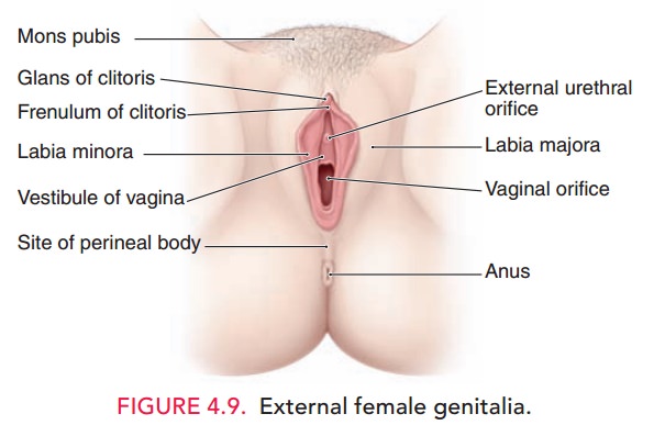
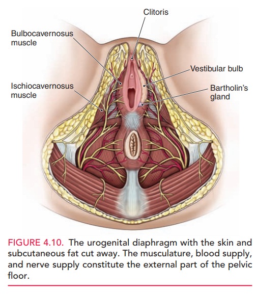
At its lower end, the vagina
traverses the urogenital diaphragm and is then surrounded by the two
bulbocaver-nosus muscles of the vulva. These muscles act as a sphincter. The hymen, a fold of mucosal-covered
connective tissue, somewhat obscures the external vaginal orifice. The hymen is
fragmented into irregular remnants with sexual activity and childbearing. The
major blood supply to the vagina is from the vaginal artery, a branch of the hypogastric artery, also known as
the internal iliac and parallel veins.
Related Topics